
Endochondral ossification
More of the epiphyseal zones are illustrated: the zone of maturation-hypertrophy-calcification lies near the top of the image; the zone of degeneration occupies the middle division; and the zone of ossification is at the bottom. 400x
![Zones of M-H-C > <p>In the zone of maturation-hypertrophy-calcification (arrow) [M-H-C], chondrocytes begin to mature, hypertrophy and secrete alkaline phosphatase into the matrix. An alkaline environment encourages the calcification of the cartilage matrix, thus prohibiting diffusion of nutrients to the chondrocytes.</p>](https://digitalhistology.org/wp-content/uploads/2025/04/Endo-9-MHC.png)
Zones of M-H-C >
In the zone of maturation-hypertrophy-calcification (arrow) [M-H-C], chondrocytes begin to mature, hypertrophy and secrete alkaline phosphatase into the matrix. An alkaline environment encourages the calcification of the cartilage matrix, thus prohibiting diffusion of nutrients to the chondrocytes.
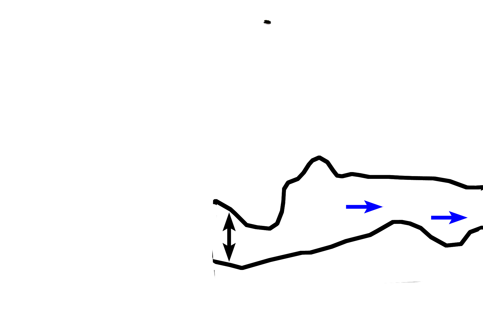
Zone of degeneration >
Having been deprived of nutrients and oxygen, chondrocytes die, leaving behind their empty lacunae. The vertical spicules of calcified cartilage (blue arrows) separate the empty lacunae and comprise the zone of degeneration.
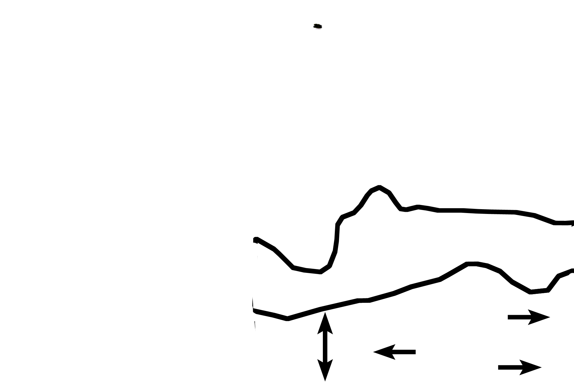
Zone of ossification >
In this zone, red-staining bone (single arrows) is deposited by osteoblasts onto calcified cartilage spicules. The calcified cartilage appears purplish-blue. Osteoblasts are carried into the marrow space along with the periosteal bud (an artery). Bone and calcified cartilage tissues are readily distinguished by their staining affinities with hematoxylin and eosin.