
Endochondral ossification
Two zone of the epiphyseal plate are shown in this image. Above the top line is the zone of proliferation; between the two lines is the zone of maturation-hypertrophy-calcification. 400x
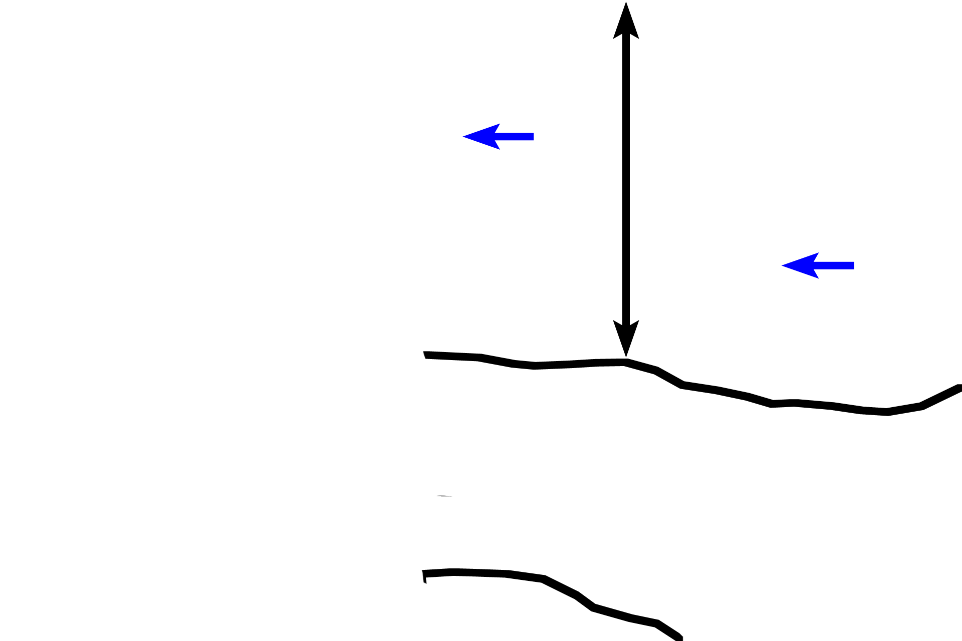
Zone of proliferation >
At the top of the image in the zone of proliferation, where chondrocytes are dividing to form linear isogenous groups of cells (blue arrows). Linear proliferation maintains the thickness of the epiphyseal plate and advances the epiphysis upwards.

Zones of M-H-C >
In this region, chondrocytes are maturing and increasing in size (hypertrophying). During this process, the cells secrete alkaline phosphatase, creating an environment conducive to calcification of the cartilage matrix. A calcified matrix prohibits nutrients from reaching the chondrocytes, and the cells die. This sequence of regressive changes forms the zone of maturation-hypertrophy-calcification.