
Vestibule
This image demonstrates several subdivisions of the middle and inner ear: stapes, oval window, vestibule with utricle, semicircular canal and duct, and cochlea. 30x
Middle ear >
The middle ear houses the ear ossicles. The stapes is the ossicle that fits into the oval window in the wall separating the middle ear from the vestibule of the inner ear. Movement of the stapes results in change of pressure in the perilymph of the inner ear, thus triggering a response in the inner ear receptors.
- Stapes
The middle ear houses the ear ossicles. The stapes is the ossicle that fits into the oval window in the wall separating the middle ear from the vestibule of the inner ear. Movement of the stapes results in change of pressure in the perilymph of the inner ear, thus triggering a response in the inner ear receptors.
- Oval window
The middle ear houses the ear ossicles. The stapes is the ossicle that fits into the oval window in the wall separating the middle ear from the vestibule of the inner ear. Movement of the stapes results in change of pressure in the perilymph of the inner ear, thus triggering a response in the inner ear receptors.
Vestibule >
The vestibule is a part of the osseous labyrinth forming a portion of the inner ear. The vestibule houses the saccule (not seen here) and utricle, components of the membranous labyrinth.
- Utricle >
This section cuts through the utricle in two locations. A thickening in the epithelial lining of the utricle forms the macula, a neuroepithelial receptor, that responds to linear acceleration and gravitational forces, thereby initiating a nerve impulse in CN VIII.
-- Macula of utricle
This section cuts through the utricle in two locations. A thickening in the epithelial lining of the utricle forms the macula, a neuroepithelial receptor, that responds to linear acceleration and gravitational forces, thereby initiating a nerve impulse in CN VIII.
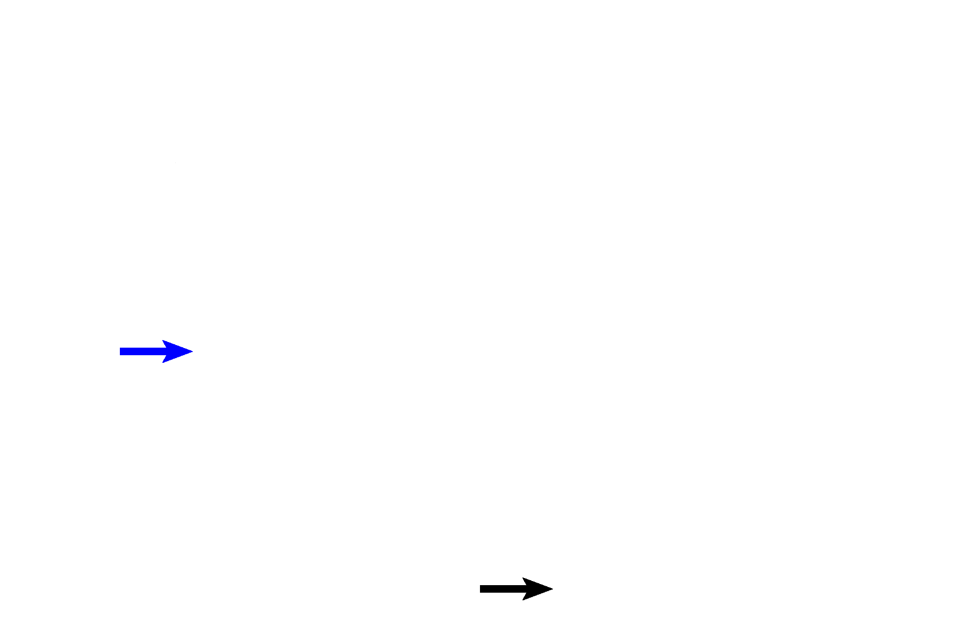
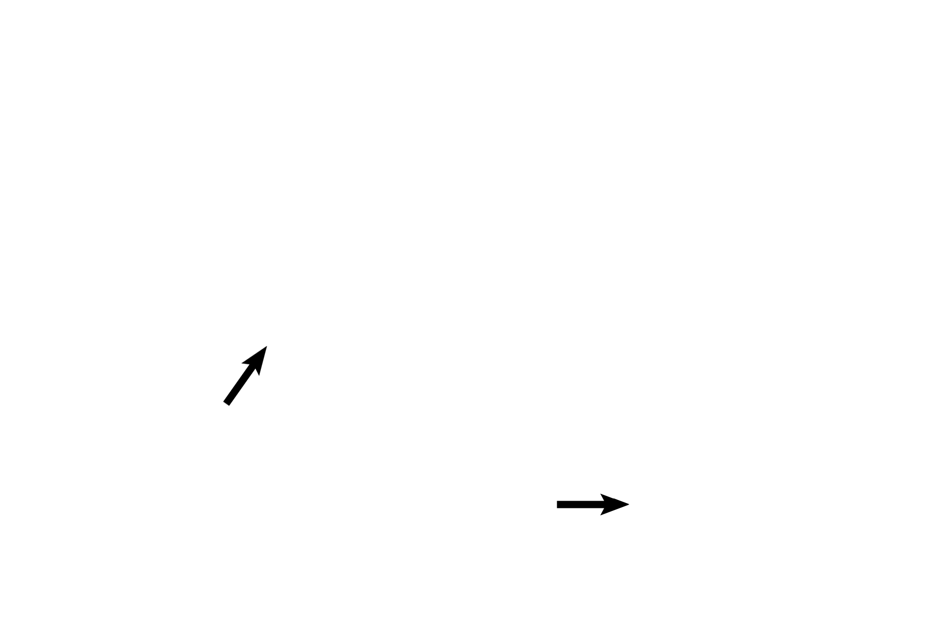
Semicircular ducts >
A portion of one (blue arrow) of the three semicircular ducts (membranous labyrinth) is sectioned at its attachment to the utricle. A second semicircular duct (black arrow) is seen a short distance from its utricular attachment.
Cochlea >
The cochlear duct (membranous labyrinth) with its organ of Corti, the receptor for audition or hearing, is encased in the cochlea, part of the osseous labyrinth.

- Cochlear duct
The cochlear duct (membranous labyrinth) with its organ of Corti, the receptor for audition or hearing, is encased in the cochlea, part of the osseous labyrinth.
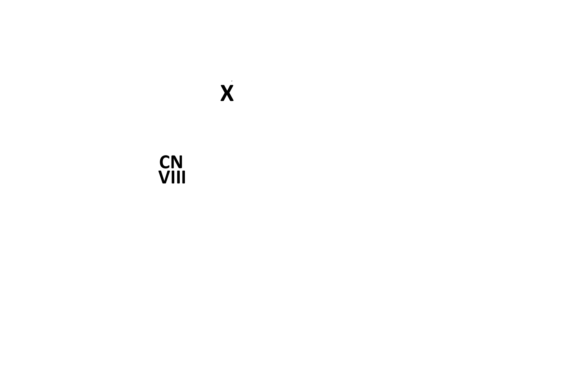
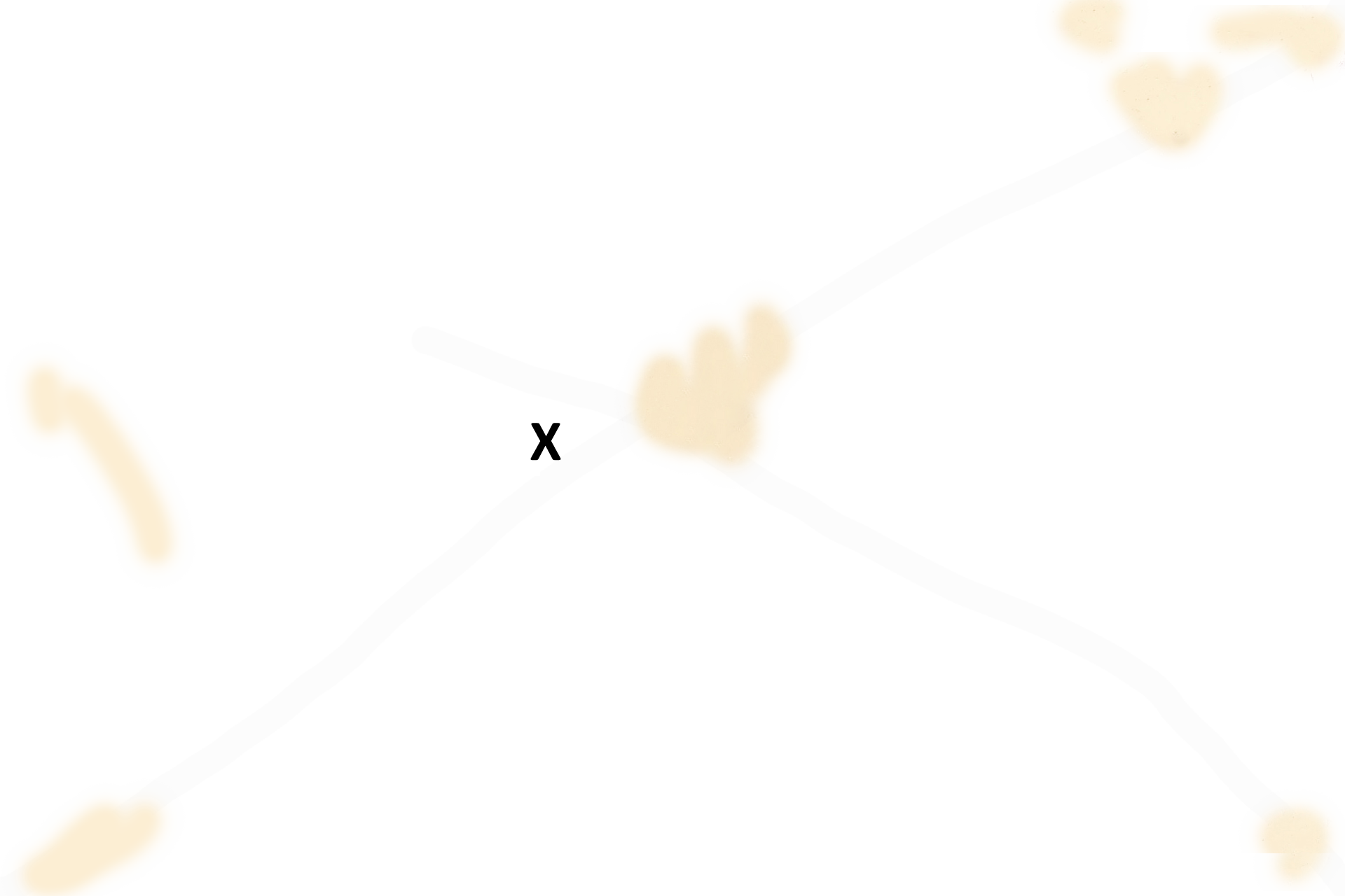
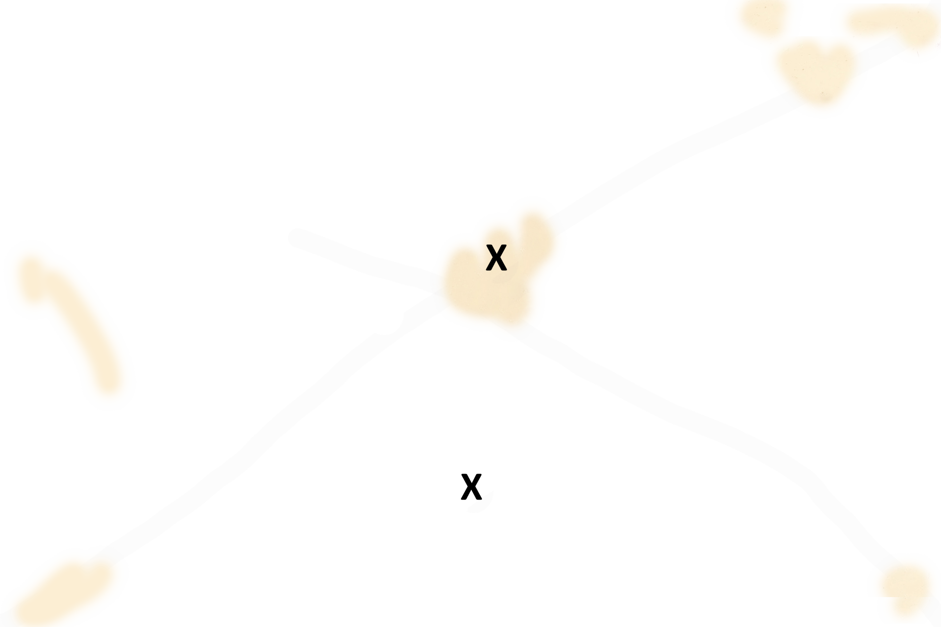
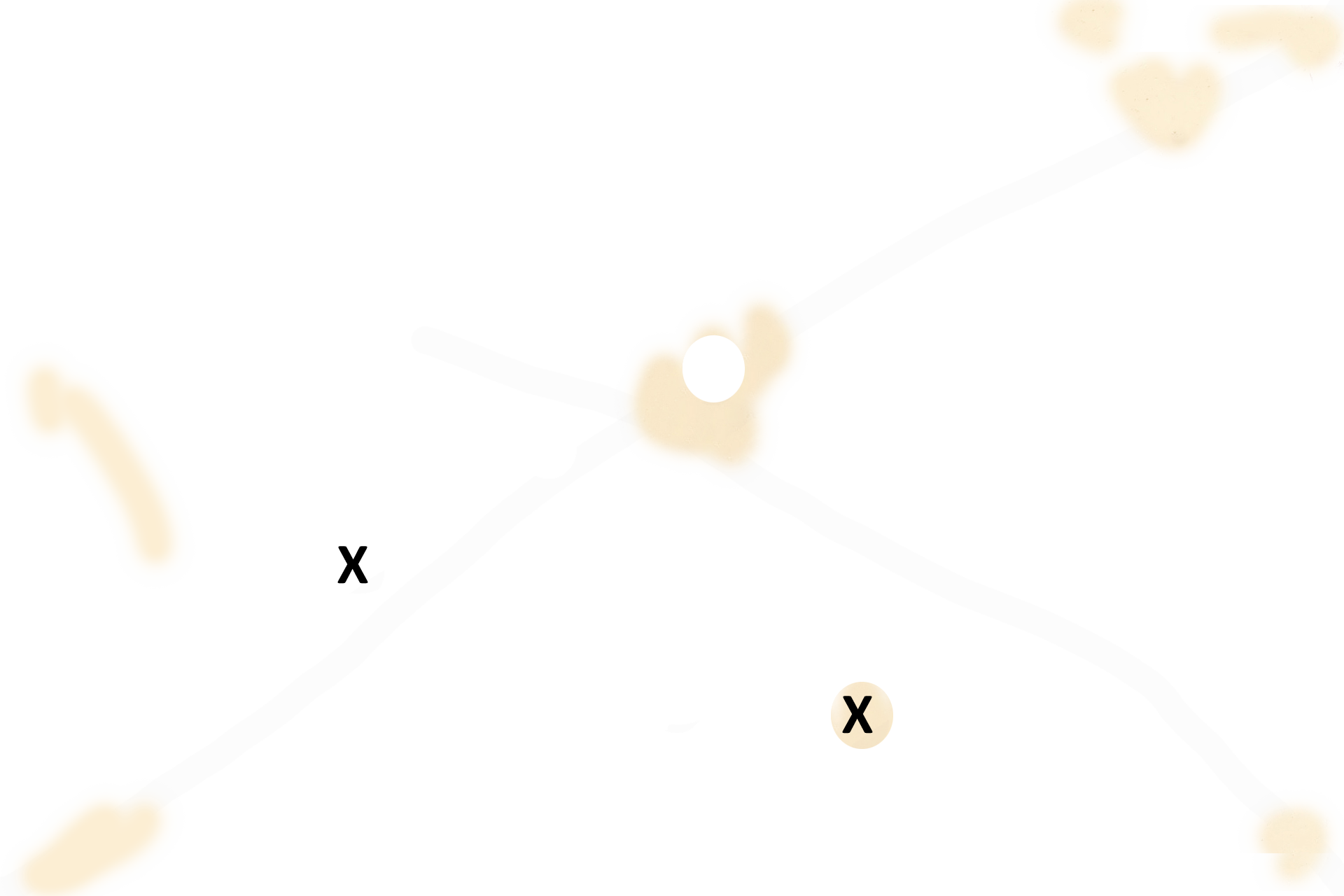
Tensor tympani and vestibulocochlear nerve >
The tensor tympani muscle (X) modulates excessively loud sounds by pulling the malleus away from the tympanic membrane. Cranial nerve VIII, the vestibulocochlear, transmits impulses from the maculae and christae ampullares to the brain.