
Vestibule
This vestibule, with associated semicircular canals and cochlea, is from a non-human source. Within the vestibule are the utricle and the saccule, portions of the membranous labyrinth. Vestibular functions, giving a sense of body position, are provided by receptors in the utricle, saccule, and semicircular ducts. The vestibular portion of cranial nerve VIII innervates these receptors. 30x
Plane of section
This vestibule, with associated semicircular canals, is from a non-human source. Within the vestibule are the utricle and the saccule, portions of the membranous labyrinth. Vestibular functions, giving a sense of body position, are provided by receptors in the utricle, saccule, and semicircular ducts. The vestibular portion of cranial nerve VIII innervates these receptors. 30x
Temporal bone >
The petrous portion of the temporal bone outlines the spaces of the inner ear that form the osseous labyrinth.
Vestibule >
The vestibule is the large perilymph-filled space of the osseous labyrinth. The vestibule contains two components of the membranous labyrinth, the utricle and the saccule.
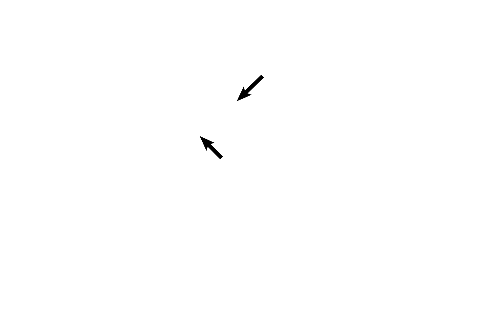
- Utricle and saccule >
The utricle (blue arrow and outline) is the larger of the two membranous structures in the vestibule. The smaller portion of the membranous labyrinth is the saccule (black arrows). Each structure possesses a macula, a thickening in its floor that is a neuroepithelial receptor serving vestibular functions.
-- Maculae >
Each macula (black arrows) contains hair cells that possess stereocilia and a cilium, embedded in a gelatinous layer. Otoliths are suspended at the top of the gel. Changes in gravity and linear motion displace the otoliths, which deflect the stereocilia and cilium, thereby initiating an impulse in CN VIII.
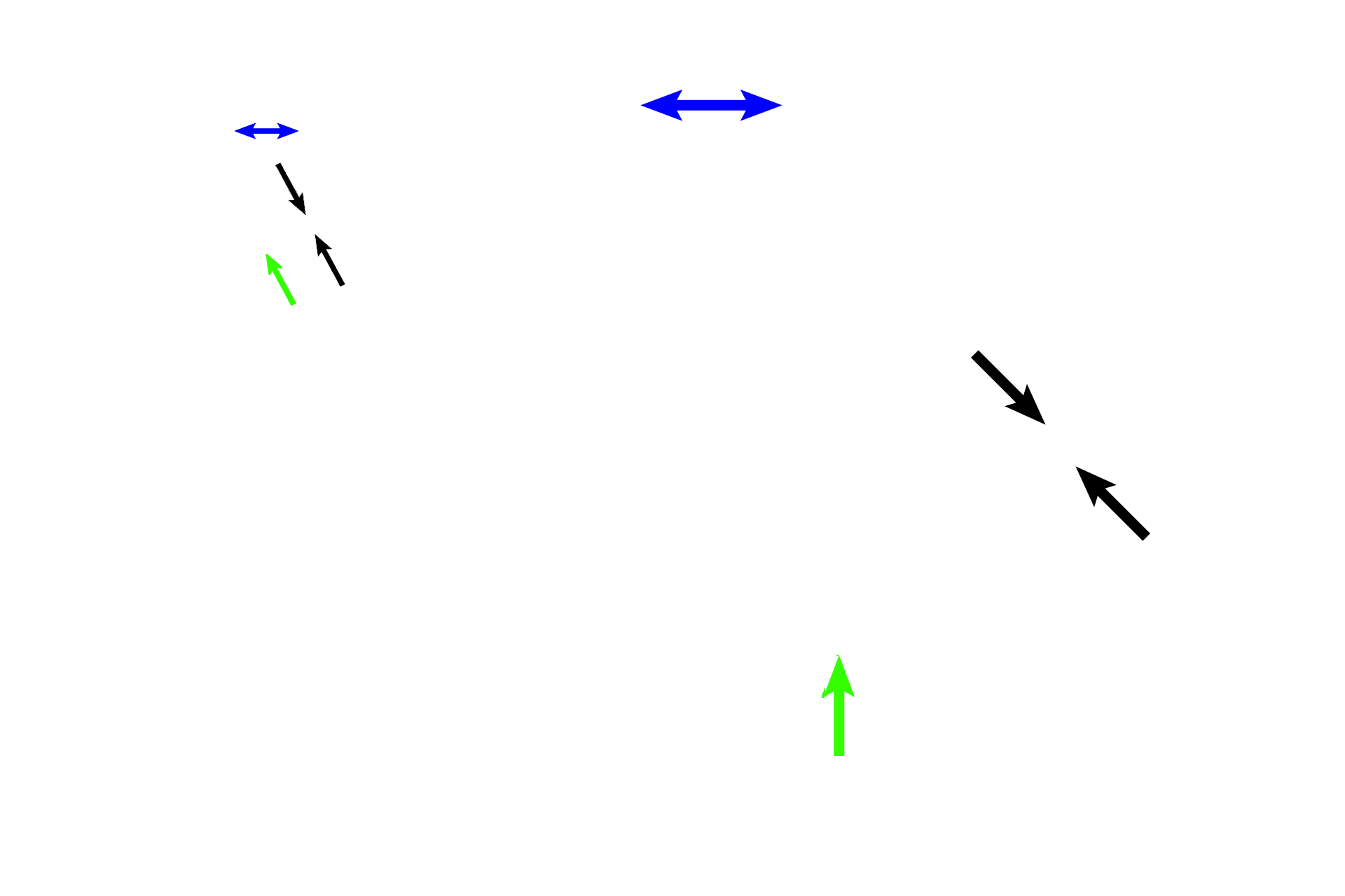
Semicircular canals >
Portions of the three semicircular canals, components of the osseous labyrinth, indicate their ring shapes and their attachments to the vestibule. The attachment of one tube is shown with its enlargement, the ampulla (blue arrows). A section of a second canal (black arrows) is visible, and the third canal (green arrows) is shown just at its attachment with the vestibule.
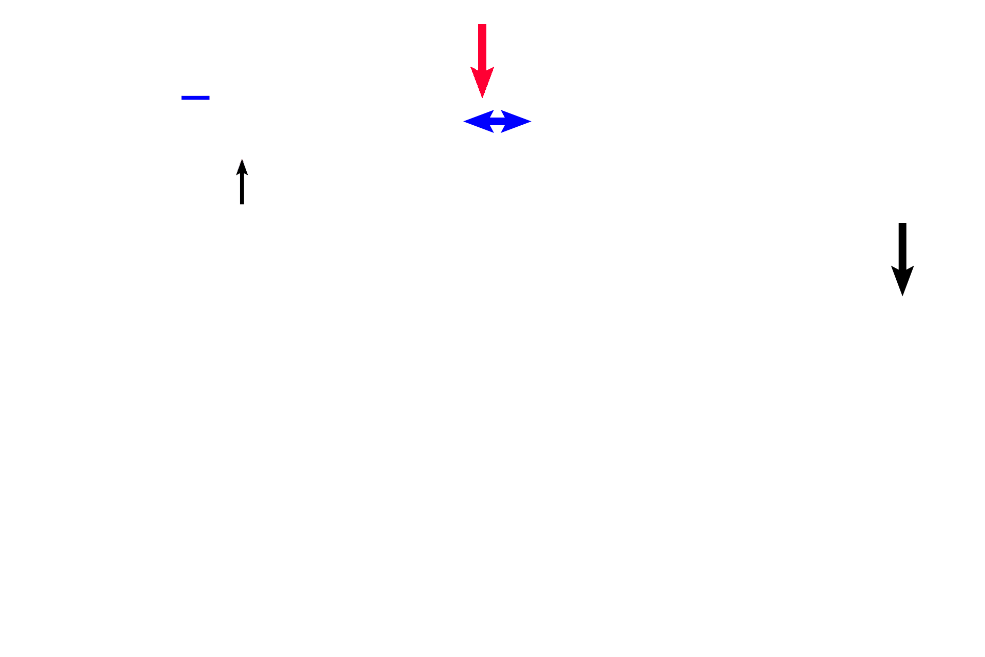
- Semicircular ducts >
Semicircular ducts (black arrows), are portions of the membranous labyrinth that are suspended in each semicircular canal and that are attached to the utricle. An enlargement at one attachment of each duct forms the ampulla (blue arrow and line). The crista ampullaris (red arrow) is the receptor located in each ampulla.
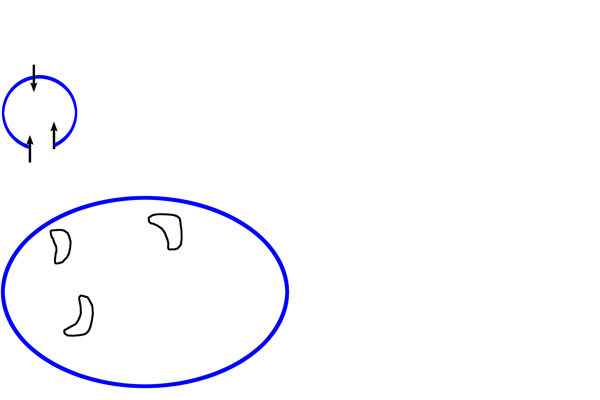
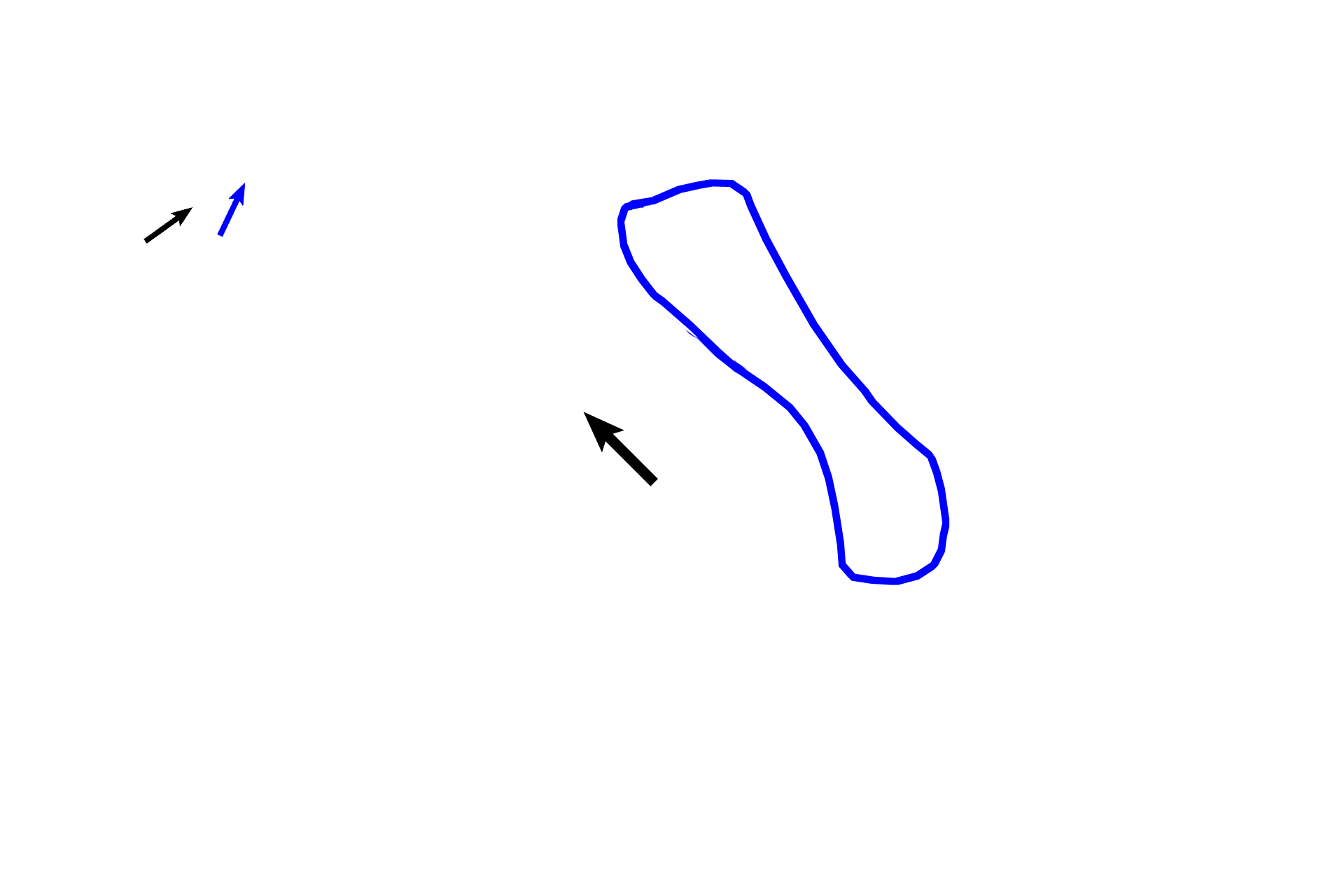
Cochlea >
The cochlea (blue outlines), a portion of the osseous labyrinth, is attached to the vestibular portion of the inner ear. The cochlear duct (black arrows and outlines), a portion of the membranous labyrinth, divides the cochlea into thirds and houses the organ of Corti, the receptor for hearing.

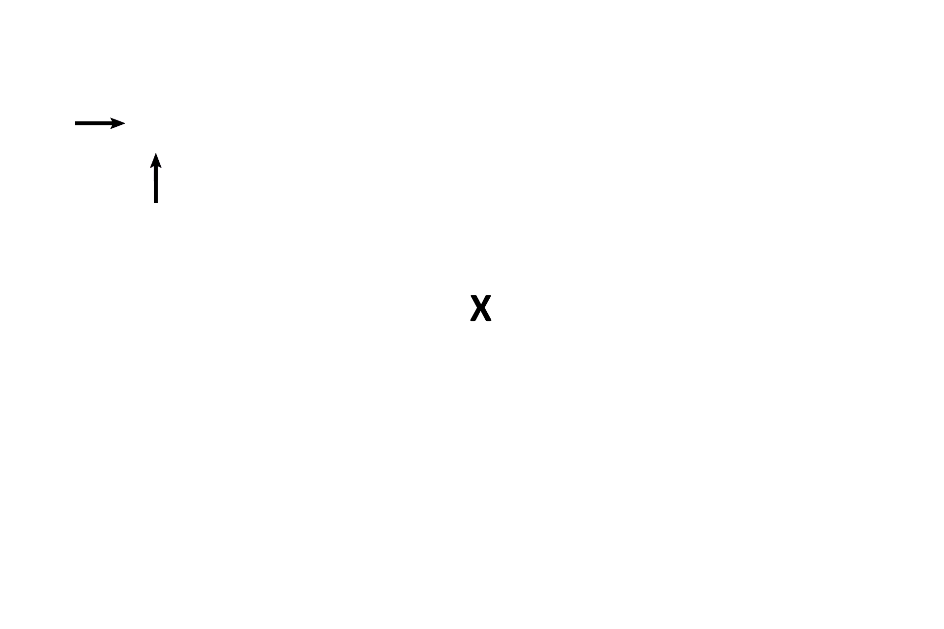
Middle ear and brain >
The cavity of the middle ear is indicated by the “X.” A section of brain is also visible (Y).