
Basic neural function
Stimuli are perceived from the internal or external environment and conveyed into the CNS via sensory neurons whose cell bodies are located in a spinal or a cranial ganglion. This input is processed by associative neurons within the CNS and a response is elicited via a motor neuron to an appropriate effector, e.g., skeletal muscle, as illustrated here.
CNS: Brain and spinal cord >
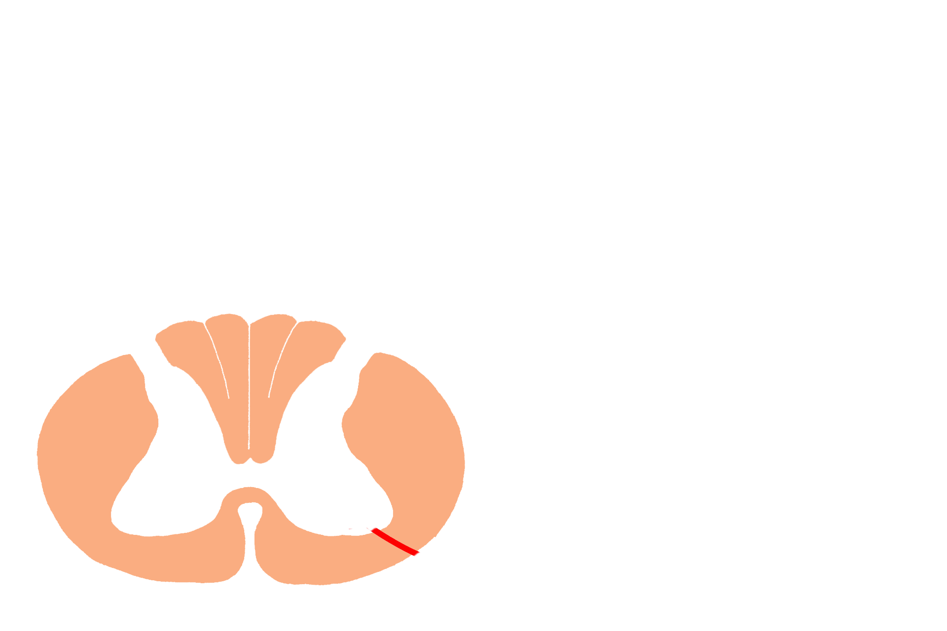
The central nervous system (CNS) consists of the brain and spinal cord, seen here. Both subdivisions have areas where nerve cell bodies are located (grey matter), and areas where axon tracts predominate (white matter). The spinal cord has a central, H-shaped area of grey matter and a peripheral zone of white matter (shaded in orange), containing axons traveling to or from higher and lower CNS centers.
PNS >
The peripheral nervous system (PNS, shaded in orange) consists of cranial nerves, spinal nerves, sensory ganglia of both cranial and spinal nerves, autonomic motor ganglia and nerves.

- Spinal nerve >
Attached to the spinal cord at regular intervals are spinal nerves, carrying information into and from the spinal cord. Each spinal nerve has a dorsal root and a ventral root.

-- Dorsal root >
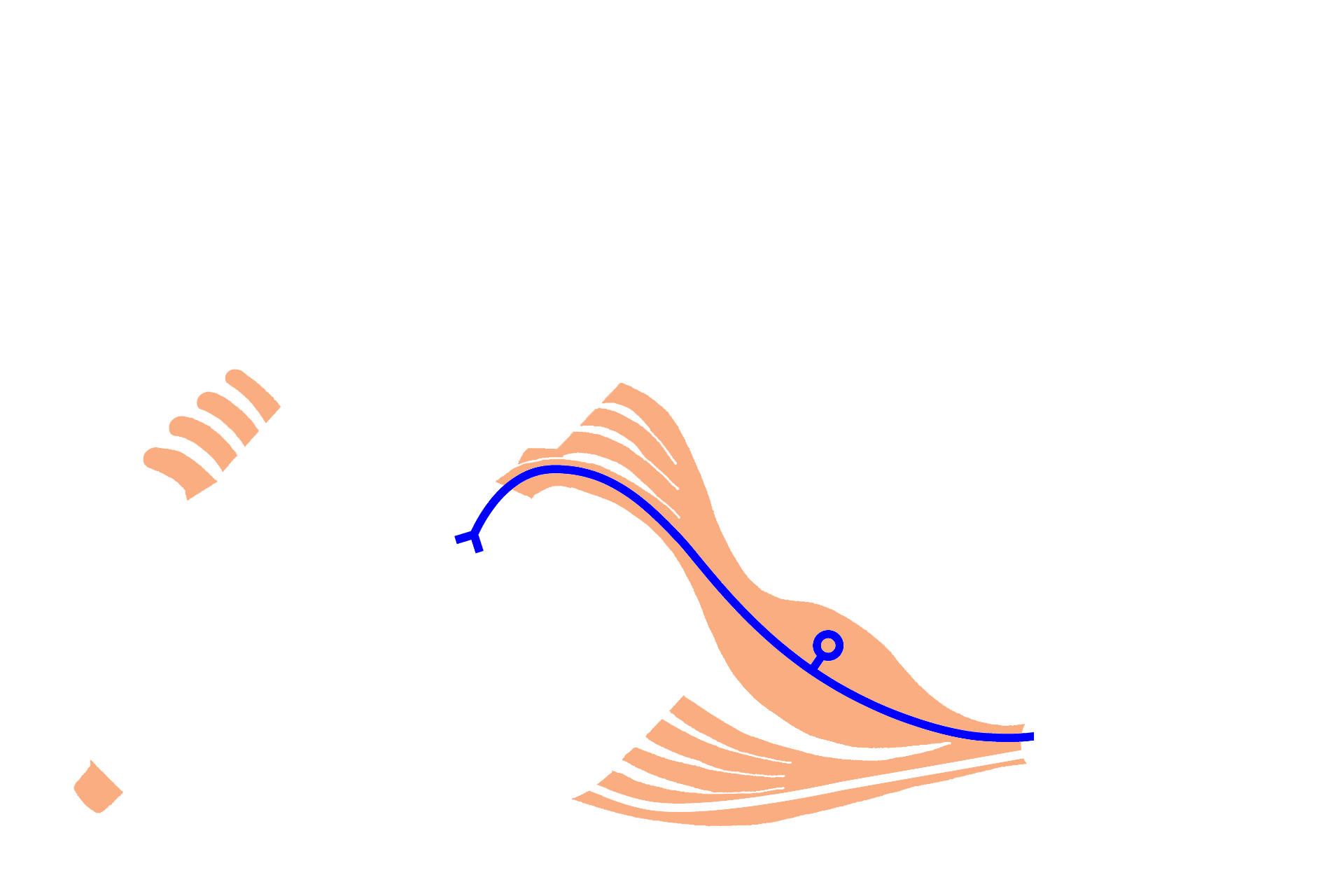
The dorsal root carries sensory information, via pseudounipolar neurons (in blue), into the spinal cord from receptors in the internal or external environment. Cell bodies of these neurons are located in a sensory ganglion on the dorsal root called a dorsal root ganglion. A ganglion is an accumulation of nerve cell bodies in the peripheral nervous system.

-- Dorsal root ganglion >
The dorsal root carries sensory information, via pseudounipolar neurons (in blue), into the spinal cord from receptors in the internal or external environment. Cell bodies of these neurons are located in a sensory ganglion on the dorsal root called a dorsal root ganglion (green oval). A ganglion is an accumulation of nerve cell bodies in the peripheral nervous system.

-- Ventral root >
The ventral root of a spinal nerve carries motor information via multipolar neurons (in red) from the CNS to effector structures, such as muscles, glands, or other neurons. The nerve cell body for each multipolar neuron is located in the grey matter of the spinal cord; the axon of each neuron exits via the ventral root.

Receptor >
Receptors (such as free nerve endings, Meissner’s corpuscles or Pacinian corpuscles), illustrated here in the skin, perceive changes in the environment. These changes are transmitted into the CNS via a process of a sensory, pseudounipolar neuron.

Sensory neurons >
Sensory information is carried into the CNS by pseudounipolar neurons (in blue). These sensory neurons begin at the receptor and have their cell bodies located either in a dorsal root ganglion (as illustrated here) or in a sensory ganglion of a cranial nerve. The central process of each neuron terminates in the CNS.

Interneurons (association neurons) >
Association neurons are multipolar neurons located entirely within the CNS. These neurons integrate information they receive from multiple sources, including neurons located elsewhere in the CNS and in the PNS, such as these pseudounipolar neurons. In the reflex arc illustrated here, this processed information is conveyed to effector neurons which stimulate muscles or glands.

Motor neuron >
Motor neurons are multipolar with their cell bodies predominately located in the CNS. Axons of these motor, multipolar neurons travel via the ventral root of a spinal nerve to an effector organ.

Effector organ >
Effector structures in the periphery respond to input from a multipolar, motor neuron whose axon travels in the ventral root. This nerve stimulus results in the contraction of a muscle (such as the skeletal muscle illustrated here), the secretion of a gland, or the stimulation of another neuron.

Integration >
Spinal cord activity does not remain confined to a single segment of the spinal cord. Activity can be relayed upward or downward a few spinal cord segments or it may reach higher cognitive centers in the brain.