
Multipolar neuron
This pyramidal neuron in the cerebral cortex is an example of a multipolar neuron. It has been stained by the Golgi silver impregnation method which clearly labels the cell with a black precipitate that covers the entire neuron. 400x

Cell body >
The cell body of pyramidal neurons is roughly triangular or pyramid-shaped. Two additional neurons, slightly out of the plane of focus, are also visible.
Dendrites >
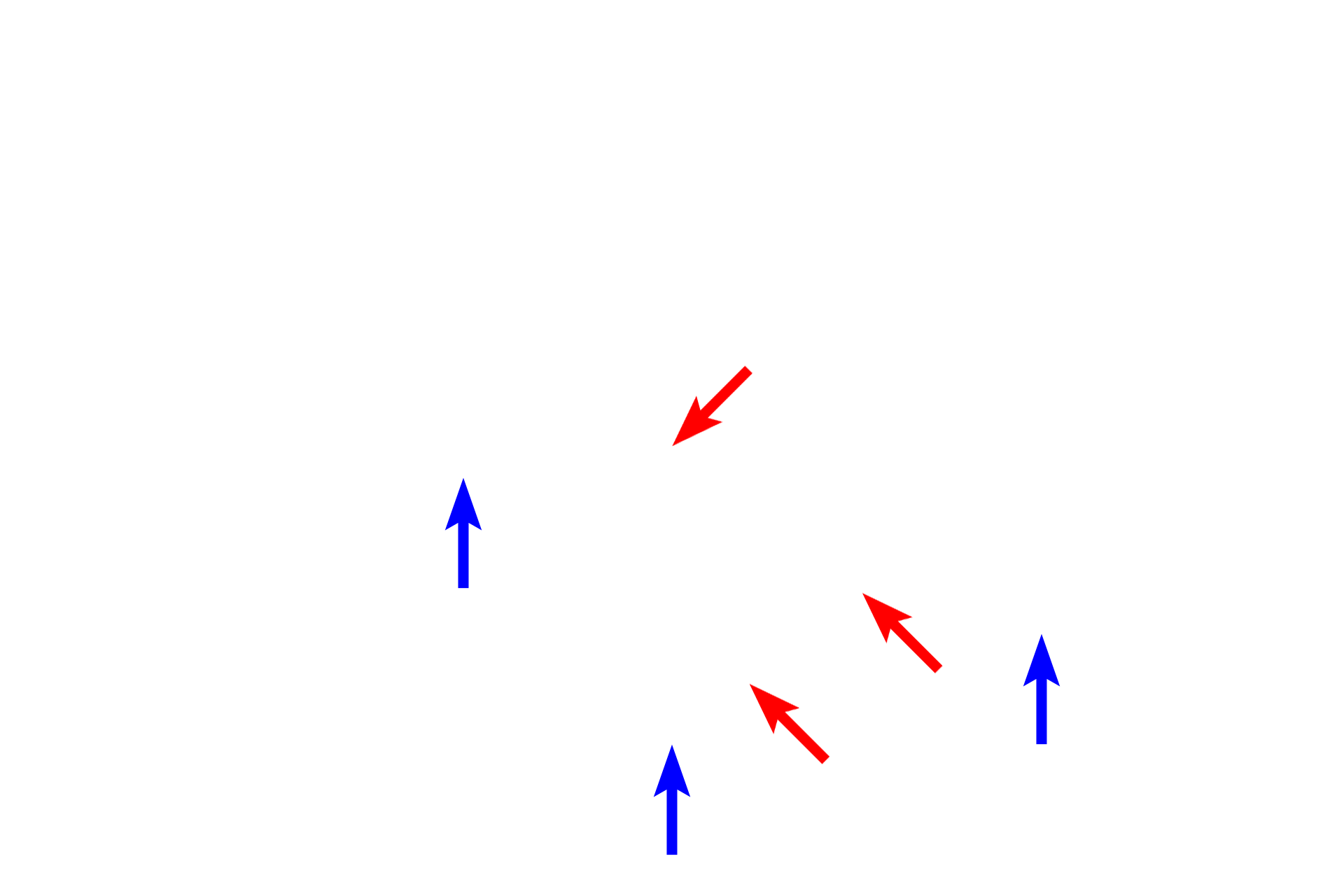
Three major dendrites (red arrows) emanate from the cell body, two from the base of the soma and one from the apex. These major dendrites branch repeatedly (blue arrows) and most display dendritic spines

Dendritic spines >
Dendritic spines are small (0.1 – 0.8 microns) protrusions of the dendrite plasma membrane and are highly dynamic structures that remodel with synaptic activity. The majority of excitatory input to dendrites occurs on dendritic spines.

Capillary
Dendritic spines are small (0.1 – 0.8 microns) protrusions of the dendrite plasma membrane and are highly dynamic structures that remodel with synaptic activity. The majority of excitatory input to dendrites occurs on dendritic spines.

Image source >
This images was taken of a slide on the Virtual Microscopy Laboratory website (www.histologyguide.com).