
Fibrocartilage
This image shows the transition zone between the dense regular connective tissue of the muscle tendon (left) to fibrocartilage (middle). The collagen fibers eventually connect to bone on the right. To best transmit the force of muscle contraction, the collagen bundles of the tendon are arranged in a parallel manner (dense, regular connective tissue). The tendon gradually transitions to fibrocartilage, where cartilage cells and their rubbery matrix occupy the spaces between the fibers. This construction resists the compressional forces at the insertion while still transmitting the force of contraction to the bone. 400x
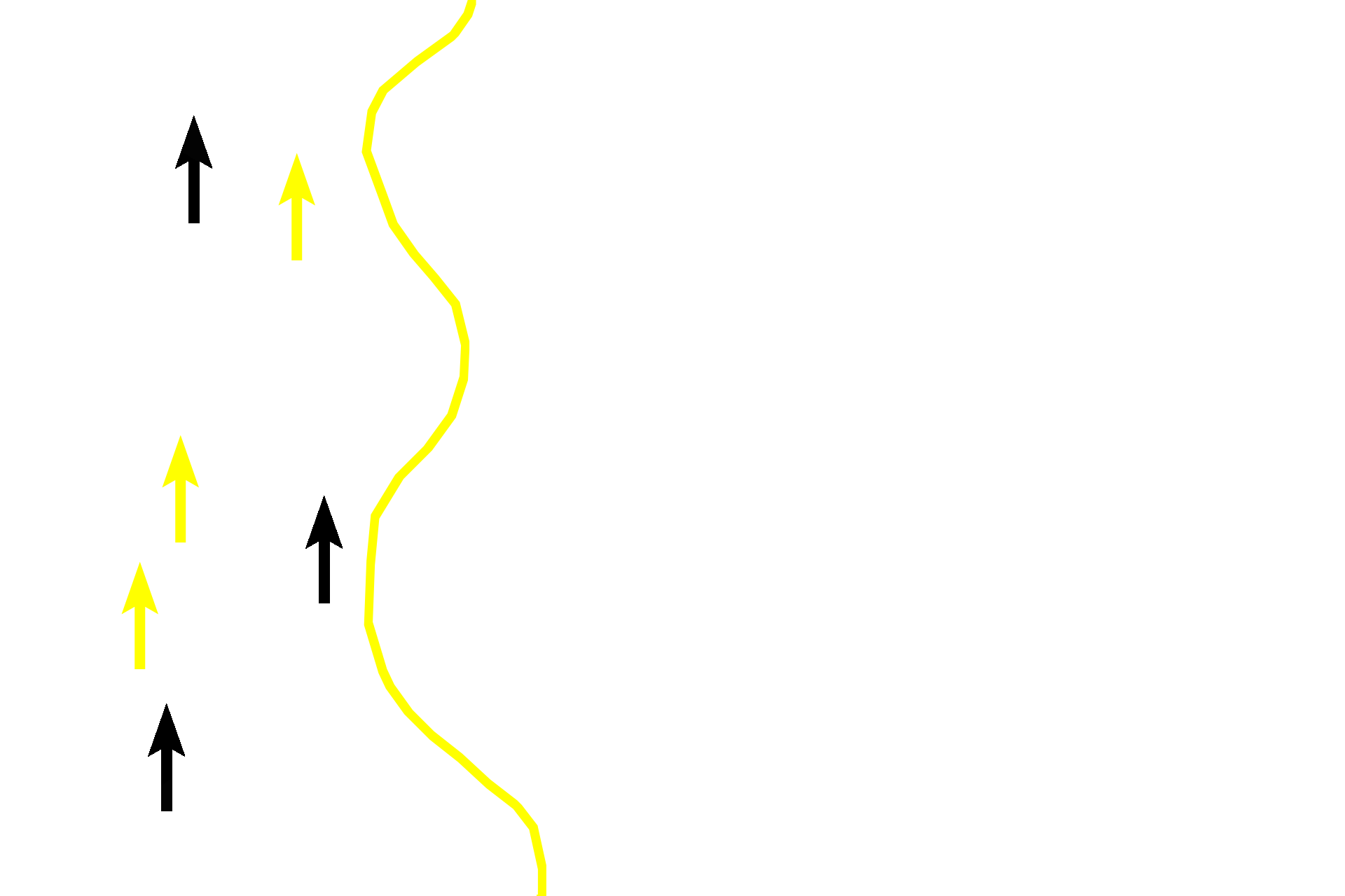
Dense regular connective tissue >
The dense regular connective tissue forming the tendon is seen to the left of the yellow line. The flattened, inactive fibroblast nuclei (yellow arrows) are arranged linearly between bundles of red collagen fibers. Small slivers of fibrocartilage can be seen extending between the collagen bundles (black arrows).
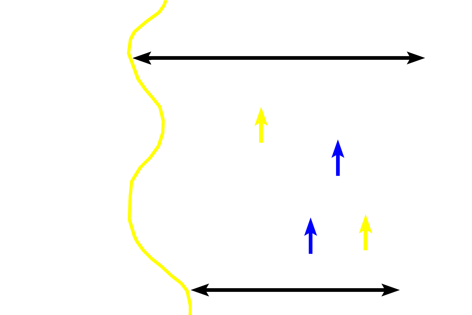
Fibrocartilage >
Coursing through this fibrocartilage are pink/red collagen fibers (yellow arrows) separated by rows of chondrocytes and their pale-staining matrix (blue arrows). The fibers continue from the tendon through the fibrocartilage to embed in bone, thus firmly anchoring the tendon to the bone. The presence of the fibrocartilage between the fibers resists the compressional forces generated by the muscle contraction. Abundant ground substance causes fibrocartilage to stain more blue than does tendon or bone.

Bone >
The continuation of the collagen fibers from the fibrocartilage insert into the both to transmit the force of muscle contraction.
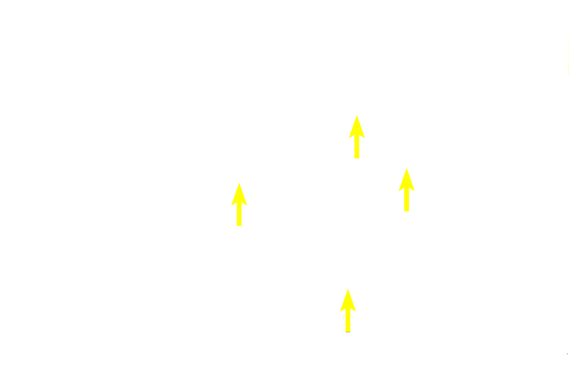
Chondrocytes >
Chondrocytes of fibrocartilage are located between parallel bundles of fibers, as are fibroblasts in tendon. Chondrocytes are spherical because they are surrounded by a firm ground substance that resists compressional forces exerted by the muscle-tendon complex. No perichondrium is present because fibrocartilage lacks a free surface.
Next Image
The next image is similar to that outlined by the rectangle.