
Bone the organ
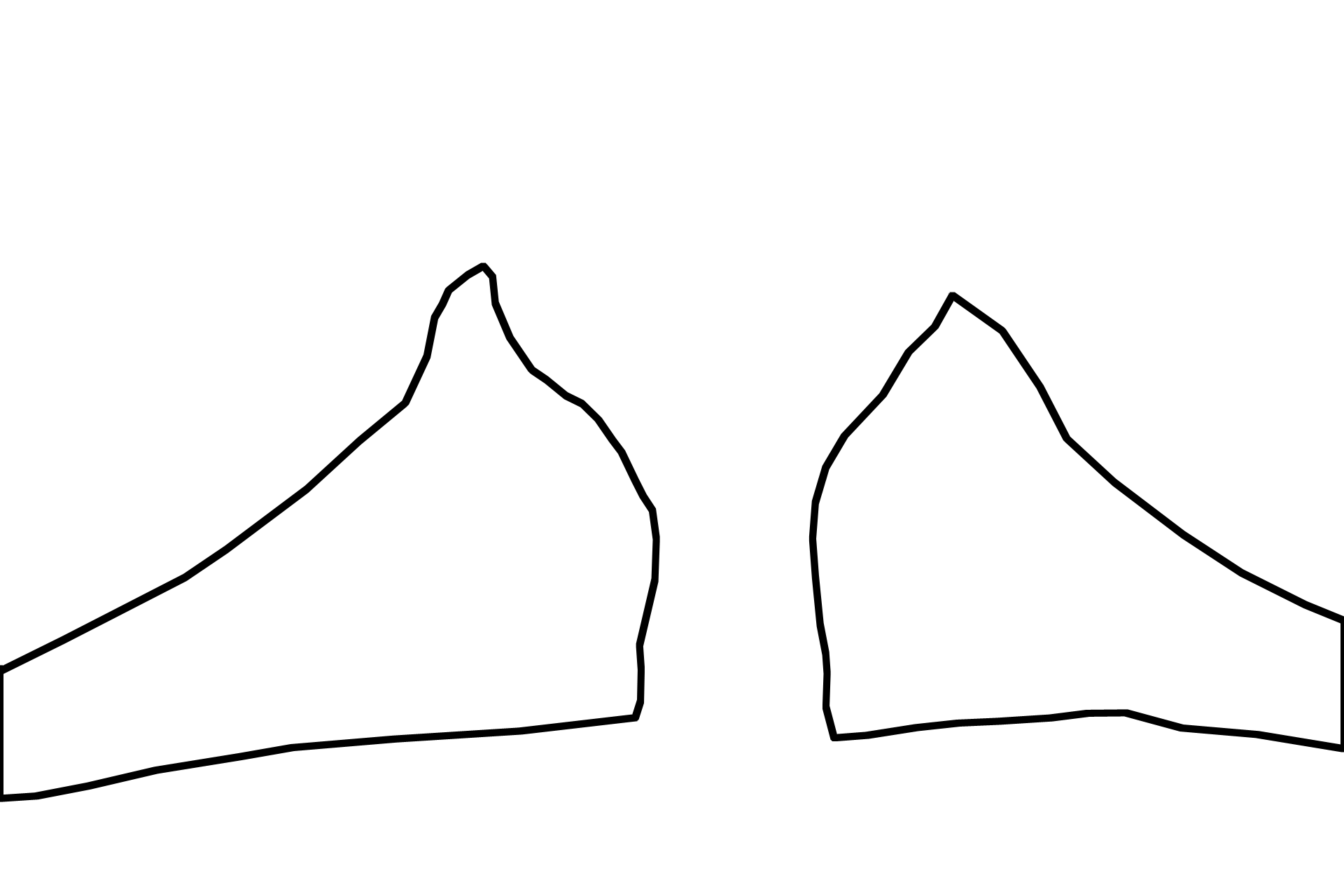
The organ, bone, consists of the tissue bone, its surface coverings, marrow, and nerve and blood supplies. In this image two wedges of pubic bones are separated by a centrally located pubic symphysis of hyaline cartilage. The feathery, red strands radiating from the bone are skeletal muscles. 10x
Bone >
A wedge-shaped, red-staining, pubic bone extends from each lateral surface of the central, blue-staining cartilage. Bone stains red because of its abundant collagen and minimal ground substance. As is typical, the periphery of this bone is formed of compact bone while its center is filled with spongy bone alternating with islands of marrow.
Compact bone
A wedge-shaped, red-staining, pubic bone extends from each lateral surface of the central, blue-staining cartilage. Bone stains red because of its abundant collagen and minimal ground substance. As is typical, the periphery of this bone is formed of compact bone while its center is filled with spongy bone alternating with islands of marrow.
Spongy bone
A wedge-shaped, red-staining, pubic bone extends from each lateral surface of the central, blue-staining cartilage. Bone stains red because of its abundant collagen and minimal ground substance. As is typical, the periphery of this bone is formed of compact bone while its center is filled with spongy bone alternating with islands of marrow.

Marrow
A wedge-shaped, red-staining, pubic bone extends from each lateral surface of the central, blue-staining cartilage. Bone stains red because of its abundant collagen and minimal ground substance. As is typical, the periphery of this bone is formed of compact bone while its center is filled with spongy bone alternating with islands of marrow.

Hyaline cartilage >
Hyaline cartilage, forming the fetal pubic symphysis, stains blue because of the abundant, negatively charged ground substance it contains. This tissue will be replaced with a pad of fibrocartilage by adulthood.
Muscle >
Skeletal muscle inserts into the compact bone forming the bony periphery.

Next Image
The next image is similar to the area outlined by the rectangle.