
Nasal cavities
The nasal cavities are shown in the diagram (left) and in a histological image (right). The blue shaded areas in the diagram represent spaces within the nasal cavity, pharynx, larynx and trachea. The image on the right shows the nasal cavity in a frontal section of the head. The symmetry in this image was digitally generated for illustration purposes from a single hemisection. 10x
Area shown on the right
The nasal cavities are shown in the diagram (left) and in a histological image (right). The blue shaded areas in the diagram represent spaces within the nasal cavity, pharynx, larynx and trachea. The image on the right shows the nasal cavity in a frontal section of the head. The symmetry in this image was digitally generated for illustration purposes from a single hemisection. 10x

Nasal cavities >
The nasal cavities are extrapulmonary passages that are part of the conducting portion of the respiratory system. The two nasal cavities are separated by the nasal septum and their lateral walls are formed by the bony nasal conchae (turbinate bones). Most of the nasal cavities are lined by respiratory epithelium.

Nasal septum >
The two nasal cavities are separated by the nasal septum which is formed by hyaline cartilage in its anterior region and bone posteriorly.

Conchae (turbinates) >
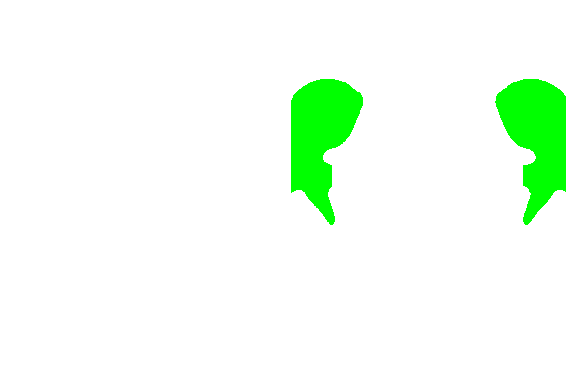
Conchae are shelves of bone that protrude into the nasal cavity. The three pairs of conchae, inferior, middle and superior, are mostly lined by respiratory mucosa that serves to warm and moisten inspired air.
Olfactory epithelium >
Highly specialized olfactory epithelium lines the upper portions of the nasal septum and the upper portions of the superior turbinate bones. Olfactory epithelium contains neurons that detect odorants in the air, transmitting that information to the brain for the perception of smell. The remainder of the nasal cavity is lined by respiratory epithelium.
Paranasal sinuses >
Paranasal sinuses are air-filled cavities within the skull bones that extend from the nasal cavities. They are lined by respiratory epithelium and serve to humidify the air and lighten the skull.
Image source >
The image on the right was taken of a slide in the University of New England College of Osteopathic Medicine slide collection. The symmetry in this image was digitally generated for illustration purposes from a single hemisection.