
Epididymis: head
The testis lies anterior to the comma-shaped epididymis. Only a portion of the head of the epididymis is present in this image. 40x

Seminiferous tubules
The testis lies anterior to the comma-shaped epididymis. Only a portion of the head of the epididymis is present in this image. 40x
Tunica albuginea
The testis lies anterior to the comma-shaped epididymis. Only a portion of the head of the epididymis is present in this image. 40x
Head of epididymis >
The head of the epididymis is composed of the efferent ducts coiled into cone shapes (coni vasculosi) and the beginning of the duct of the epididymis (not shown in this image).
- Efferent ducts >
The efferent ducts are coiled into 12-20 cone-shaped structures, the coni vasculosi, composed of the efferent ducts, as well as the connective tissue and abundant vasculature surrounding them. The apex of each cone faces the testis; each base faces the posterior surface of the epididymis.
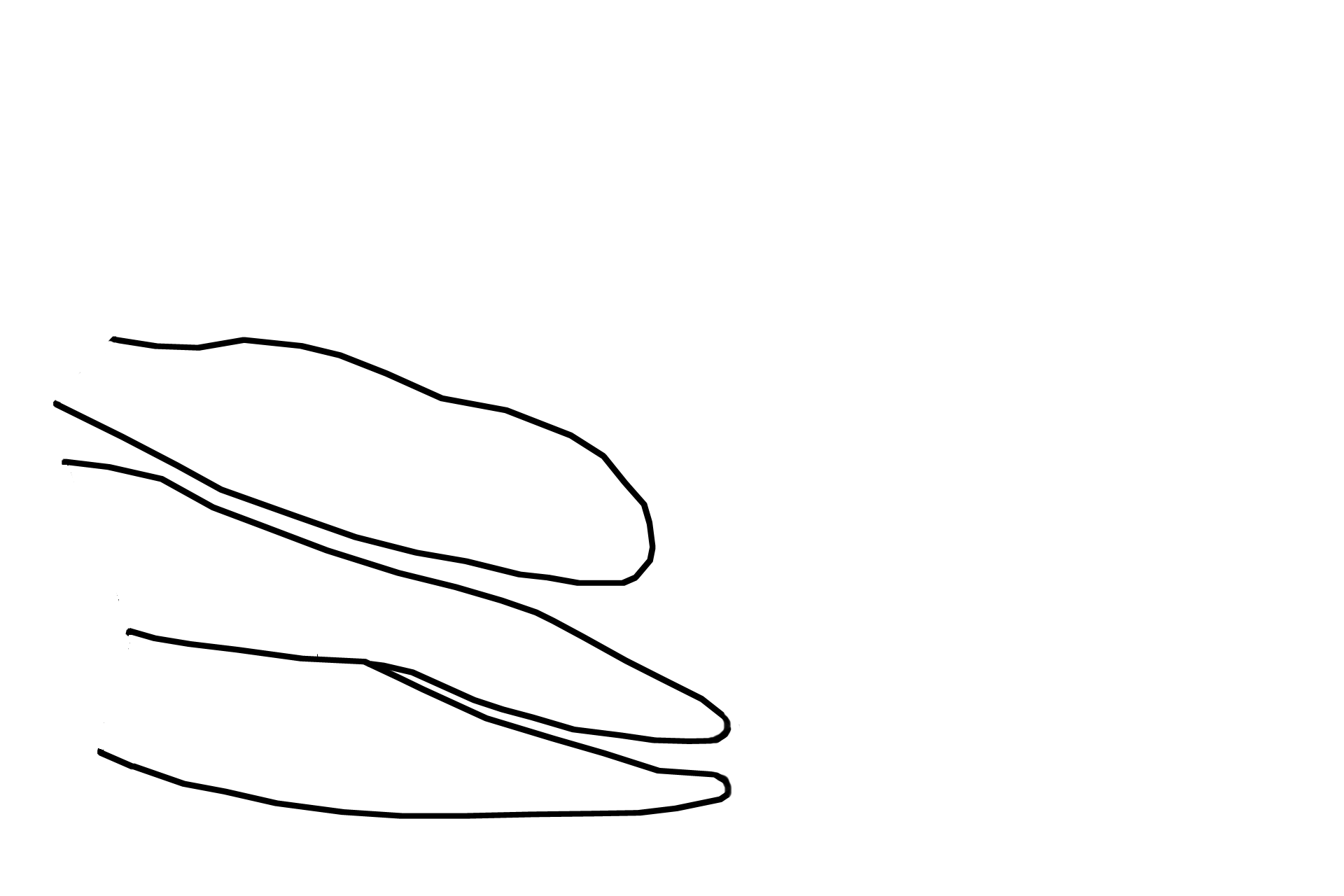
- Coni vasculosi
The efferent ducts are coiled into 12-20 cone-shaped structures, the coni vasculosi, composed of the efferent ducts, as well as the connective tissue and abundant vasculature surrounding them. The apex of each cone faces the testis; each base faces the posterior surface of the epididymis.
Next image
The next image is similar to the area outlined by the rectangle.

 PREVIOUS