
Placenta: 3rd trimester
Fetal and maternal blood in the placenta are separated by a barrier formed of fetal tissue. The barrier, separating fetal capillaries from the intervillous space, thins throughout pregnancy as the cytotrophoblastic cells decrease in number and as the syncytiotrophoblastic layer thins, allowing the numerous fetal capillaries to lie adjacent to the syncytial layer. 1000x

Intervillous space >
The intervillous space in the placenta is filled with maternal blood provided from the spurting of spiral arteries. Thus, fetal villi are bathed in maternal blood.

Floating villi >
Floating villi are those not directly attached to fetal or maternal surfaces and, thus, float free in the intervillous space. In late pregnancy, villi are composed of mucoid connective tissue with numerous fetal capillaries, a discontinuous row of cytotrophoblasts, and a syncytiotrophoblast layer with numerous microvilli to increase surface area.

- Fetal connective tissue
Floating villi are those not directly attached to fetal or maternal surfaces and, thus, float free in the intervillous space. In late pregnancy, villi are composed of mucoid connective tissue with numerous fetal capillaries, a discontinuous row of cytotrophoblasts, and a syncytiotrophoblast layer with numerous microvilli to increase surface area.

- Cytotrophoblast
Floating villi are those not directly attached to fetal or maternal surfaces and, thus, float free in the intervillous space. In late pregnancy, villi are composed of mucoid connective tissue with numerous fetal capillaries, a discontinuous row of cytotrophoblasts, and a syncytiotrophoblast layer with numerous microvilli to increase surface area.

- Syncytiotrophoblast
Floating villi are those not directly attached to fetal or maternal surfaces and, thus, float free in the intervillous space. In late pregnancy, villi are composed of mucoid connective tissue with numerous fetal capillaries, a discontinuous row of cytotrophoblasts, and a syncytiotrophoblast layer with numerous microvilli to increase surface area.

- Microvilli
Floating villi are those not directly attached to fetal or maternal surfaces and, thus, float free in the intervillous space. In late pregnancy, villi are composed of mucoid connective tissue with numerous fetal capillaries, a discontinuous row of cytotrophoblasts, and a syncytiotrophoblast layer with numerous microvilli to increase surface area.

- Fetal blood vessels >
Fetal capillaries located in floating villi are supplied by the paired umbilical arteries traveling from the fetus through the umbilical cord. These fetal capillaries anastomose to form the single umbilical vein that returns blood, rich in oxygen and nutrients, back to the fetus.
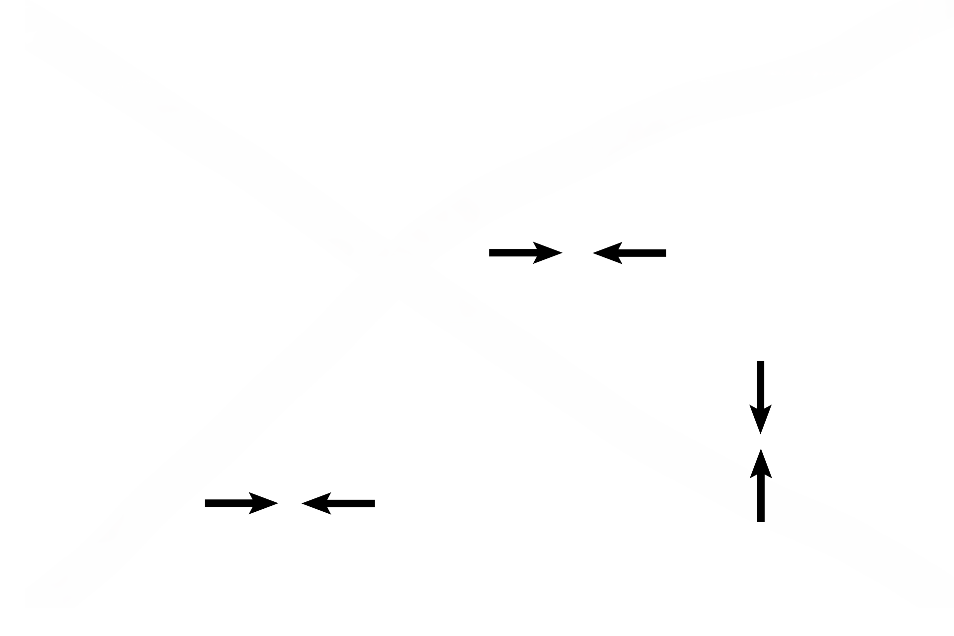
Placental barrier >
In late pregnancy the placental barrier separating fetal from maternal blood is formed by the simple squamous epithelium lining the fetal capillary, the thinned layer of syncytiotrophoblast, and their fused basement membranes. Earlier in pregnancy, the cytotrophoblastic cells and fetal connective tissue would also be included in the barrier.