
Pituitary
This image is a low magnification, midsagittal section of a human pituitary stained with hematoxylin and eosin. Note the distinct boundaries between the neurohypophyseal and adenohypophyseal tissues, indicating the different embryological origins of these subdivisions. 5x
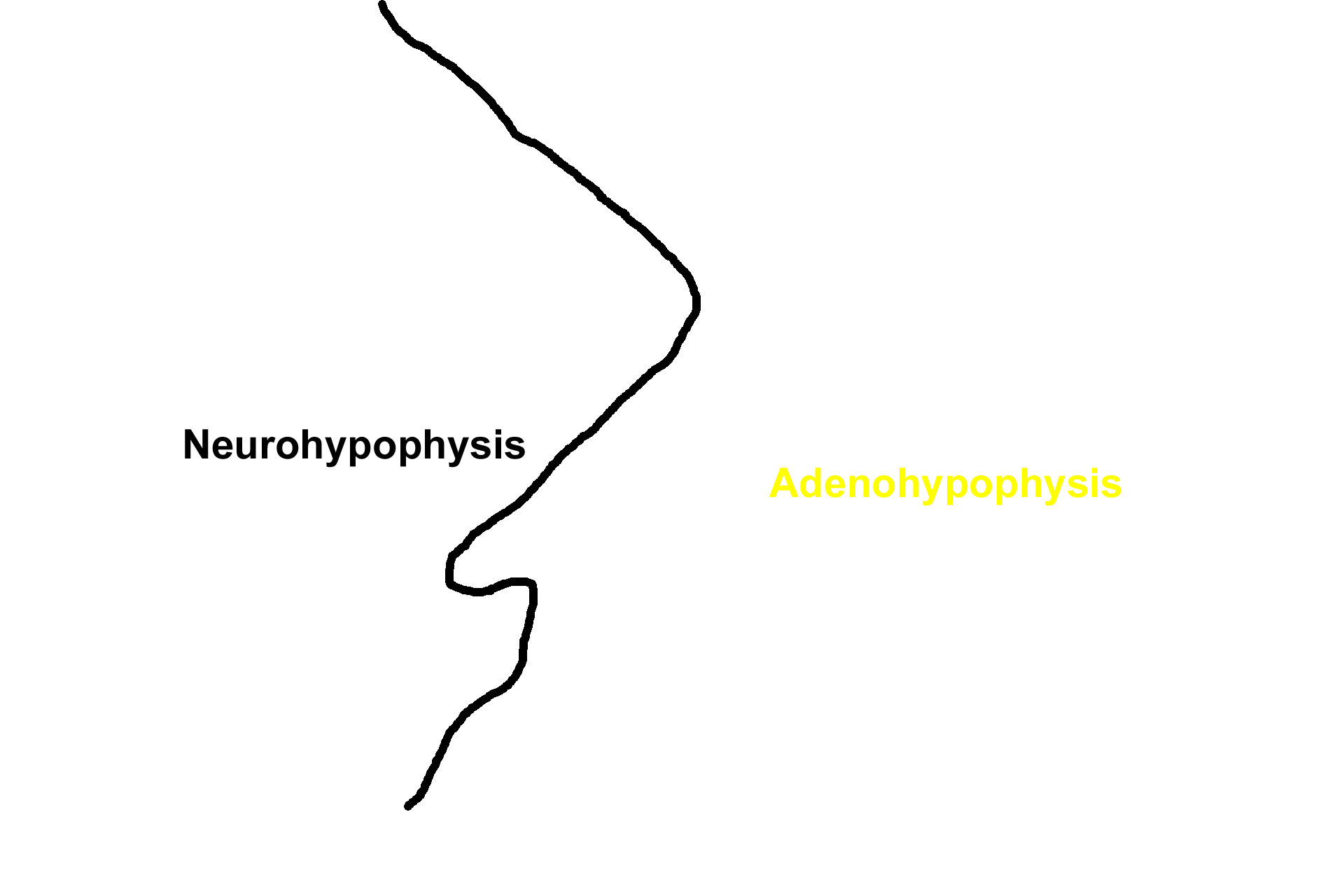
Subdivisions >
The terms neurohypophysis and adenohypophysis reflect the embryonic origin of these two subdivisions. Similar terms, posterior and anterior pituitary, are also commonly used for these subdivisions, reflecting their anatomical position.
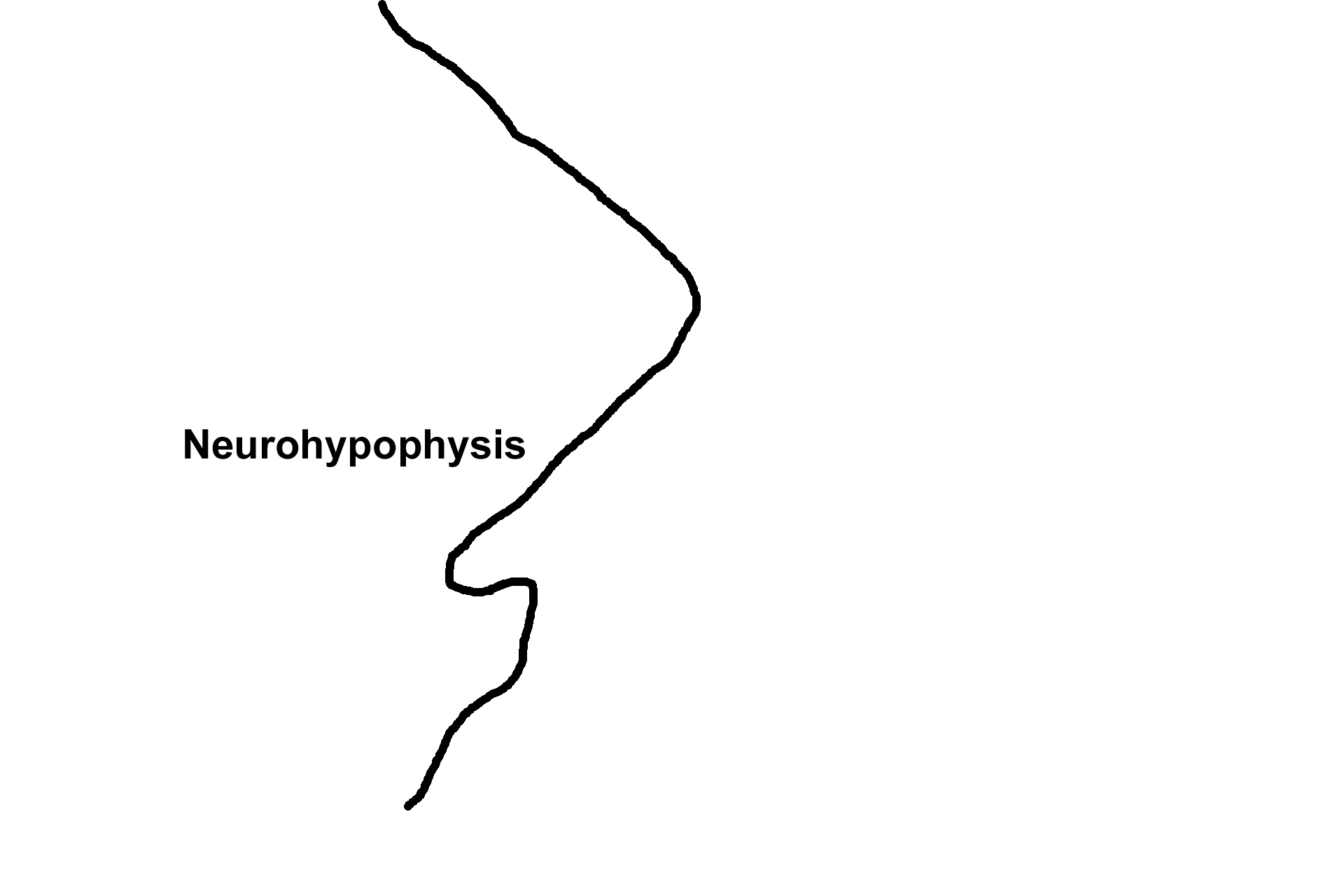
Neurohypophysis >
The neurohypophysis is derived from nervous tissue as a downgrowth from the hypothalamus of the brain and consists of the pars nervosa, the infundibulum and its continuation, the median eminence (not visible).

- Pars nervosa
The neurohypophysis is derived from nervous tissue as a downgrowth from the hypothalamus of the brain and consists of the pars nervosa, the infundibulum and its continuation, the median eminence (not visible).

- Infundibulum
The neurohypophysis is derived from nervous tissue as a downgrowth from the hypothalamus of the brain and consists of the pars nervosa, the infundibulum and its continuation, the median eminence (not visible).
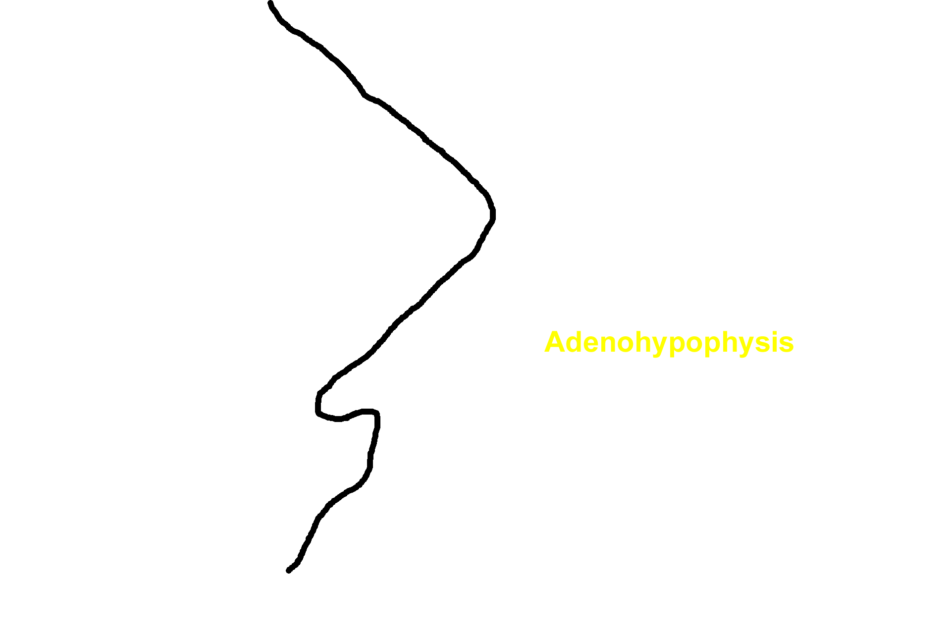
Adenohypophysis >
The adenohypophysis is derived from the epithelium that lines the roof of the developing oral cavity and consists of the pars distalis, pars tuberalis and pars intermedia.

- Pars distalis
The adenohypophysis is derived from the epithelium that lines the roof of the developing oral cavity and consists of the pars distalis, pars tuberalis and pars intermedia.

- Pars tuberalis >
The pars tuberalis forms a sleeve that surrounds the infundibulum.

- Pars intermedia >
The pars intermedia is located at the interface of the pars distalis and pars nervosa and its cells often migrate into the pars distalis.
- Rathke's pouch remnants >
The adenohypophysis originates from the oral ectoderm of the embryo as a sac-shaped structure termed Rathke’s pouch. Remnants of this pouch, typically filled with protein (colloid), are visible in the adult.

Capsule >
The entire pituitary is surrounded by a dense connective tissue capsule that is continuous with the dura mater. Only a portion of the capsule is retained in this section.

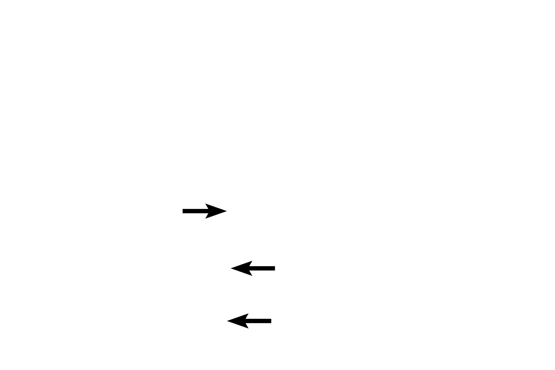
Section plane of next image >
The section plane of the next image is indicated by the horizonal line.
Image source >
This image was taken of slide in the Oklahoma State University slide collection.