
Pituitary
The pituitary gland (hypophysis) along with the associated hypothalamus of the brain, form a major regulatory system of the body. The pituitary gland is often called the master gland because it regulates numerous physiological systems, with many of its hormones targeting other endocrine organs. The hypophysis consists of two portions, the neurohypophysis and the adenohypophysis.
Hypothalamus >
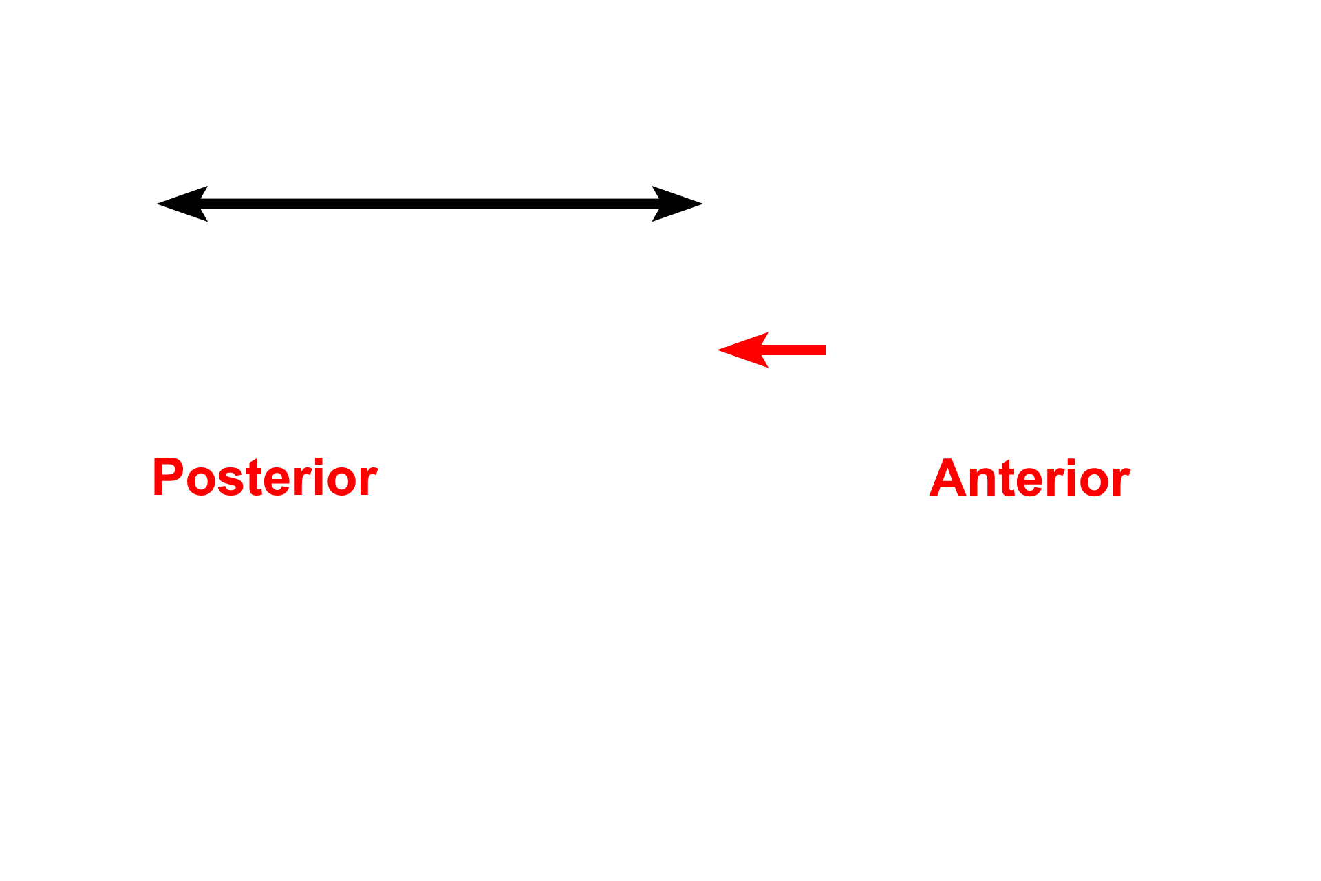
The hypothalamus (black arrow) of the brain exerts a major controlling influence over the pituitary gland. Hypothalamic neurons either directly produce neurohypophyseal hormones or indirectly stimulate or inhibit the production of adenohypophyseal hormones via a vascular connection. The optic chiasm (red arrow) is shown for reference.

Neurohypophysis >
The neurohypophysis (posterior pituitary) forms embryologically as an evagination of the hypothalamus and remains directly connected to it. The neurohypophysis consists of the pars nervosa, the infundibulum and its continuation, the median eminence. Hypothalamic neurons terminating in the neurohypophysis store and release vasopressin and oxytocin.

- Infundibulum >
The infundibulum connects the hypothalamus with the enlarged terminal portion of the neurohypophysis, the pars nervosa. The pars nervosa is sometimes referred to as the posterior lobe. The infundibulum also suspends the adenohypophysis.

- Pars nervosa
The infundibulum connects the hypothalamus with the enlarged terminal portion of the neurohypophysis, the pars nervosa. The pars nervosa is sometimes referred to as the posterior lobe. The infundibulum also suspends the adenohypophysis.
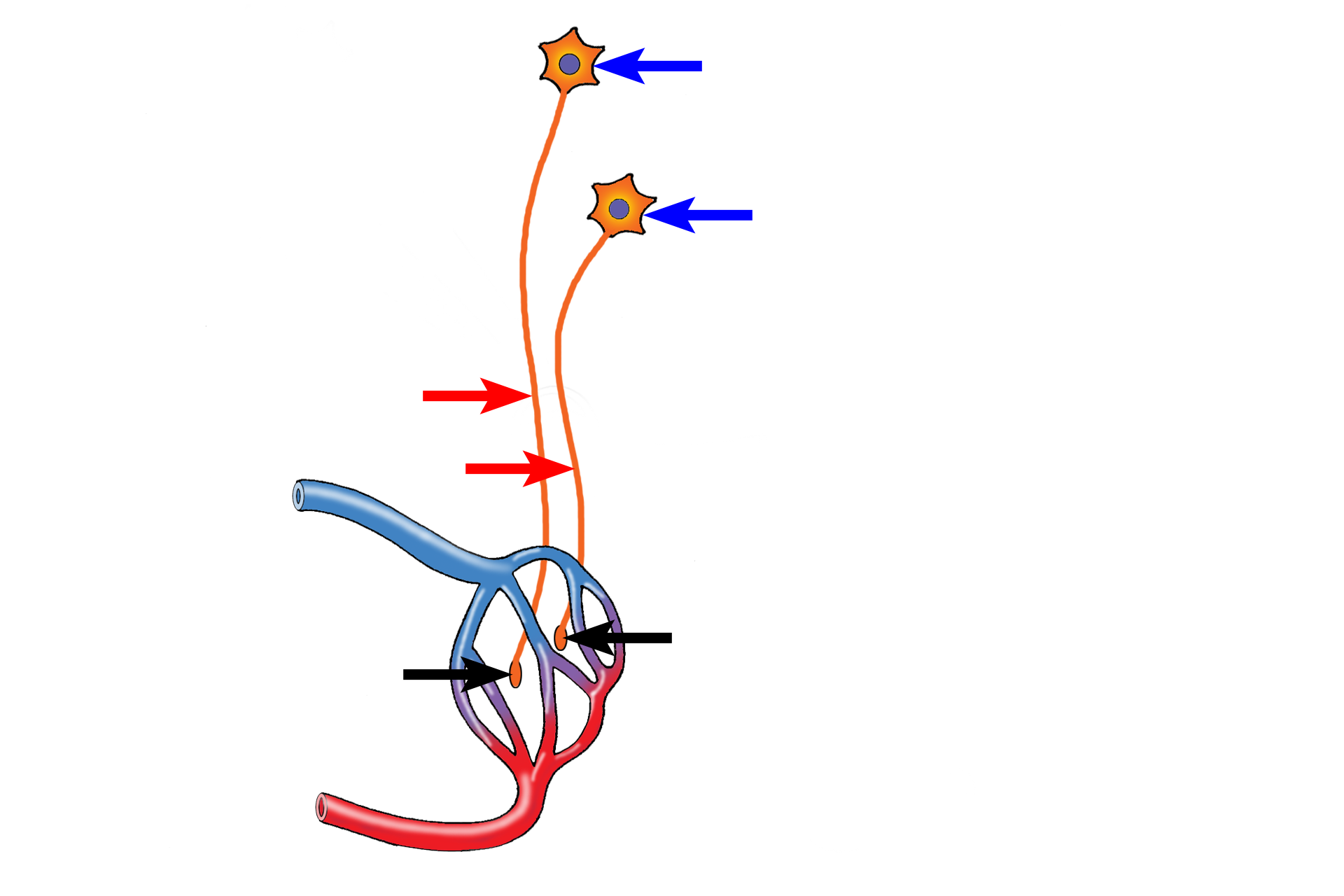
- Secretion >
Axons (red arrows) of neurons originating in supraoptic and paraventricular nuclei (blue arrows) of the hypothalamus terminate in the pars nervosa as enlargements called Herring bodies (black arrows). The hormones produced by these neurons, vasopressin and oxytocin, are stored in and released from the Herring bodies into the blood stream for distribution throughout the body.

Adenohypophysis >
The adenohypophysis (anterior pituitary) invaginates from the oral ectoderm of the embryo as a balloon-shaped structure termed Rathke’s pouch. It eventually separates from the oral cavity and comes to lie in close contact with the neurohypophysis. The synthesis and release of adenohypophyseal hormones are stimulated or inhibited by factors produced by hypothalamic neurons. The adenohypophysis consists of the pars distalis, pars tuberalis, pars intermedia and the remnants of Rathke’s pouch.

- Pars distalis >
The pars distalis is the enlarged anterior wall of Rathke’s pouch and is the largest portion of the adenohypophysis. It is sometimes referred to as the anterior lobe. The pars tuberalis extends upward from this main body of the adenohypophysis and forms an incomplete sleeve around the infundibulum.

- Pars tuberalis
The pars distalis is the enlarged anterior wall of Rathke’s pouch and is the largest portion of the adenohypophysis. It is sometimes referred to as the anterior lobe. The pars tuberalis extends upward from this main body of the adenohypophysis and forms an incomplete sleeve around the infundibulum.
- Pars intermedia >
The pars intermedia develops from the posterior wall of Rathke’s pouch. It remains small and invades the pars nervosa, making it difficult to specifically identify. In humans, the original lumen of Rathke’s pouch persists as small cysts, called the remnants of Rathke’s pouch, which separate the pars distalis from the pars intermedia. The cysts are surrounded by glandular cells of the pars intermedia.

- Remnants of Rathke's pouch
The pars intermedia develops from the posterior wall of Rathke’s pouch. It remains small and invades the pars nervosa, making it difficult to specifically identify. In humans, the original lumen of Rathke’s pouch persists as small cysts, called the remnants of Rathke’s pouch, which separate the pars distalis from the pars intermedia. The cysts are surrounded by glandular cells of the pars intermedia.
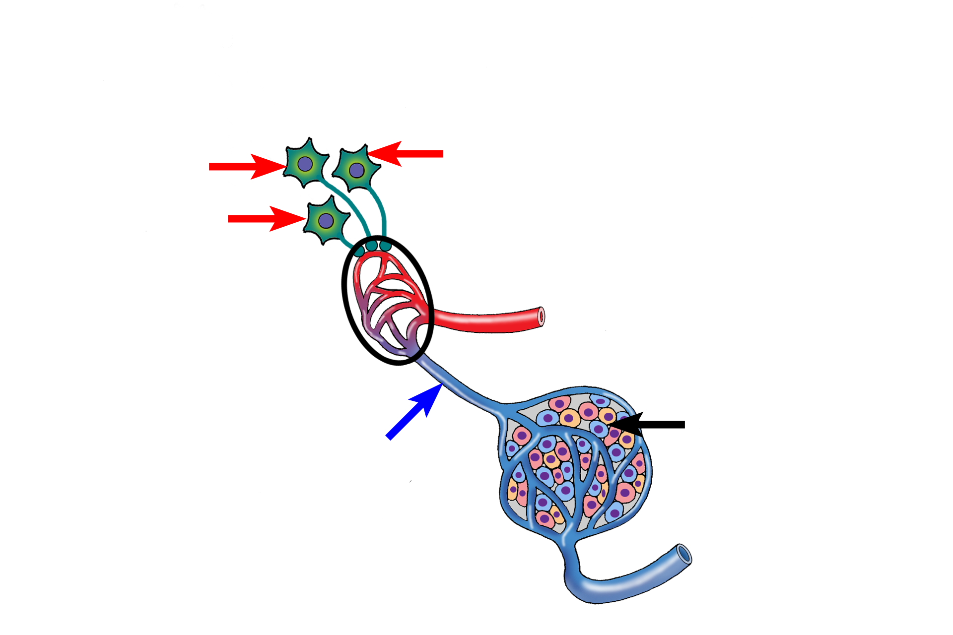
- Control of secretion >
Neurons (red arrows) in hypothalamic nuclei terminate on a capillary plexus (black circle) in the median eminence of the hypothalamus. Stimulating and inhibiting factors synthesized by these neurons are released into the capillary plexus and carried via the hypothalamo-hypophyseal portal system of vessels (blue arrow) to capillaries in the adenohypophysis, where they influence hormone secretion (black arrow).
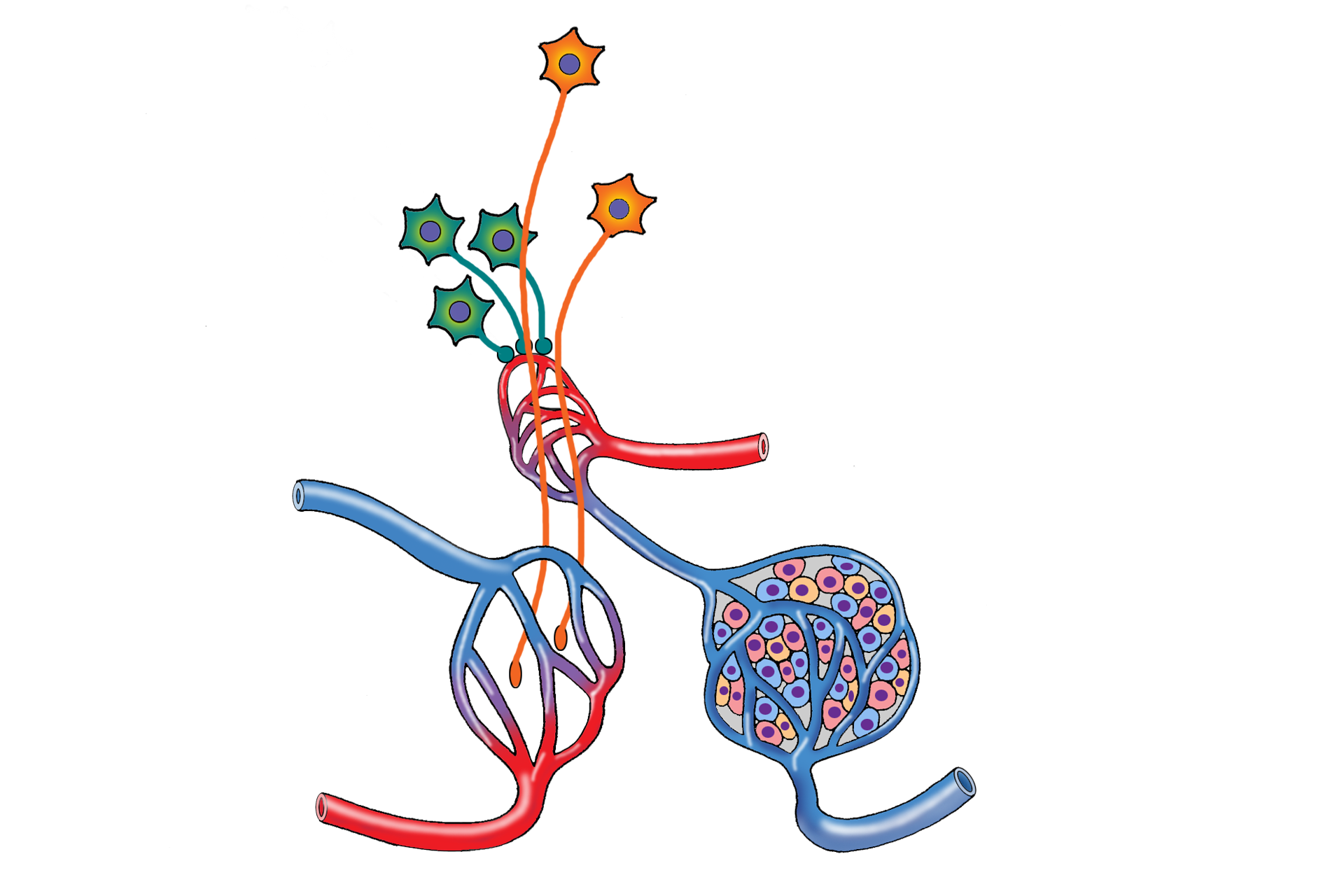
Combined secretion and control >
This composite image compares the structural and functional components of the neuro- and adenohypophyseal subdivisions, as well as their controlling mechanisms.
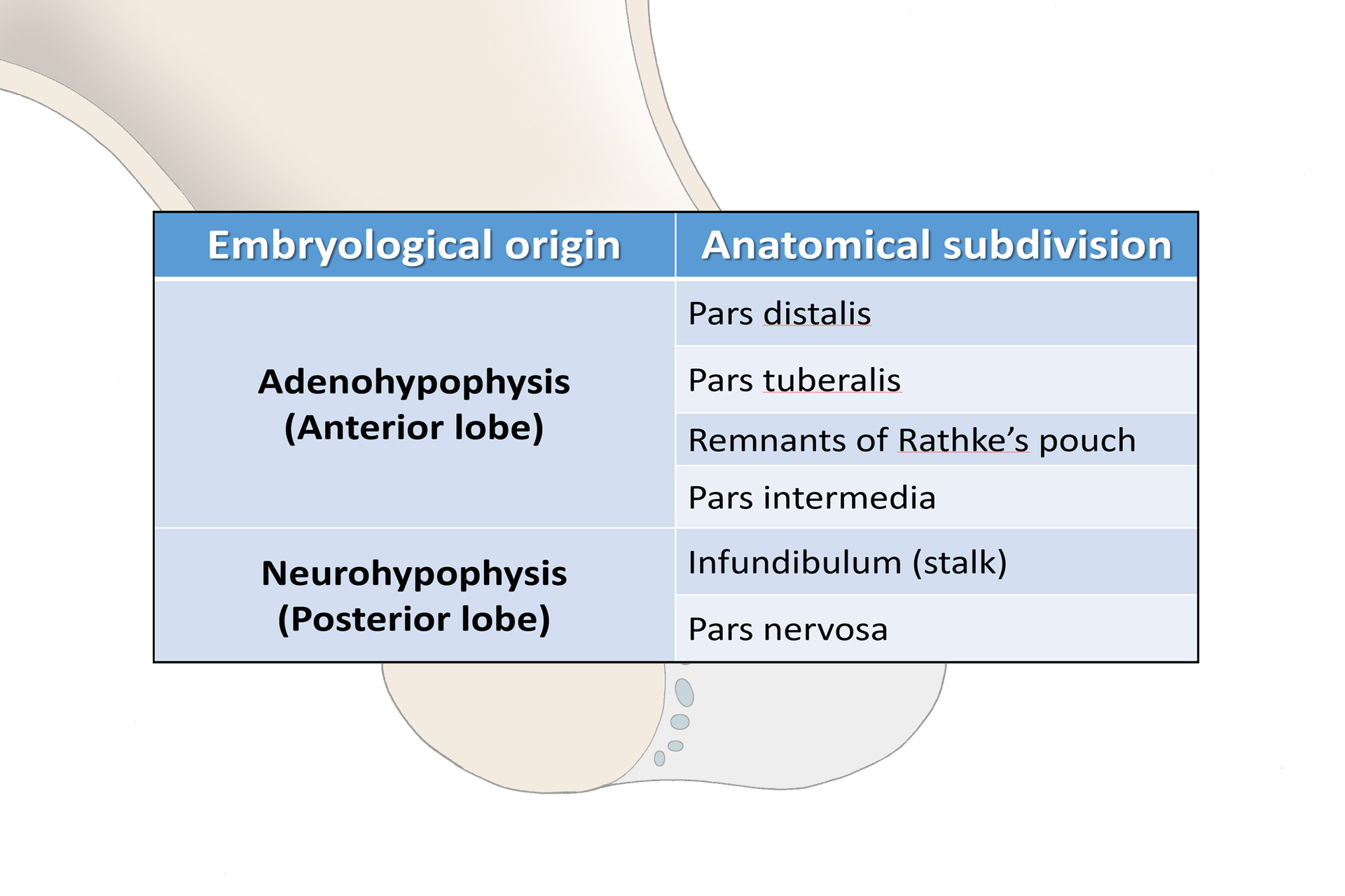
Subdivision summary >
Terminologies for the components of the pituitary gland are based on the embryological origins of the main subdivisions as well as the anatomical regions of each. In clinical reference, the term anterior pituitary is often used synonymously with pars distalis and posterior pituitary is frequently used synonymously with pars nervosa.