Fenestrated capillaries
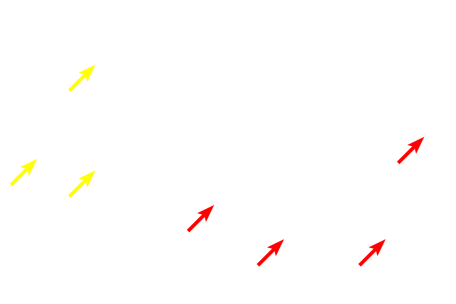
Fenestrated capillaries are present in locations, such as endocrine tissues, digestive system, and renal glomeruli, to facilitate transport through the capillary wall. On the left is a light micrograph of endocrine cells of the anterior pituitary gland. The electron micrograph on the right shows a pituitary cell adjacent to a fenestrated capillary. Fenestrations are only visible with the electron microscope. 1000x (left), 10,000x (right)

Endocrine cells
Fenestrated capillaries are present in locations, such as endocrine tissues, digestive system, and renal glomeruli, to facilitate transport through the capillary wall. On the left is a light micrograph of endocrine cells of the anterior pituitary gland. The electron micrograph on the right shows a pituitary cell adjacent to a fenestrated capillary. Fenestrations are only visible with the electron microscope. 1000x (left), 10,000x (right)

- Secretory granules
Fenestrated capillaries are present in locations, such as endocrine tissues, digestive system, and renal glomeruli, to facilitate transport through the capillary wall. On the left is a light micrograph of endocrine cells of the anterior pituitary gland. The electron micrograph on the right shows a pituitary cell adjacent to a fenestrated capillary. Fenestrations are only visible with the electron microscope. 1000x (left), 10,000x (right)

Capillaries
Fenestrated capillaries are present in locations, such as endocrine tissues, digestive system, and renal glomeruli, to facilitate transport through the capillary wall. On the left is a light micrograph of endocrine cells of the anterior pituitary gland. The electron micrograph on the right shows a pituitary cell adjacent to a fenestrated capillary. Fenestrations are only visible with the electron microscope. 1000x (left), 10,000x (right)

- Endothelium
Fenestrated capillaries are present in locations, such as endocrine tissues, digestive system, and renal glomeruli, to facilitate transport through the capillary wall. On the left is a light micrograph of endocrine cells of the anterior pituitary gland. The electron micrograph on the right shows a pituitary cell adjacent to a fenestrated capillary. Fenestrations are only visible with the electron microscope. 1000x (left), 10,000x (right)

- Fenestrae
Fenestrated capillaries are present in locations, such as endocrine tissues, digestive system, and renal glomeruli, to facilitate transport through the capillary wall. On the left is a light micrograph of endocrine cells of the anterior pituitary gland. The electron micrograph on the right shows a pituitary cell adjacent to a fenestrated capillary. Fenestrations are only visible with the electron microscope. 1000x (left), 10,000x (right)

- Pericyte process >
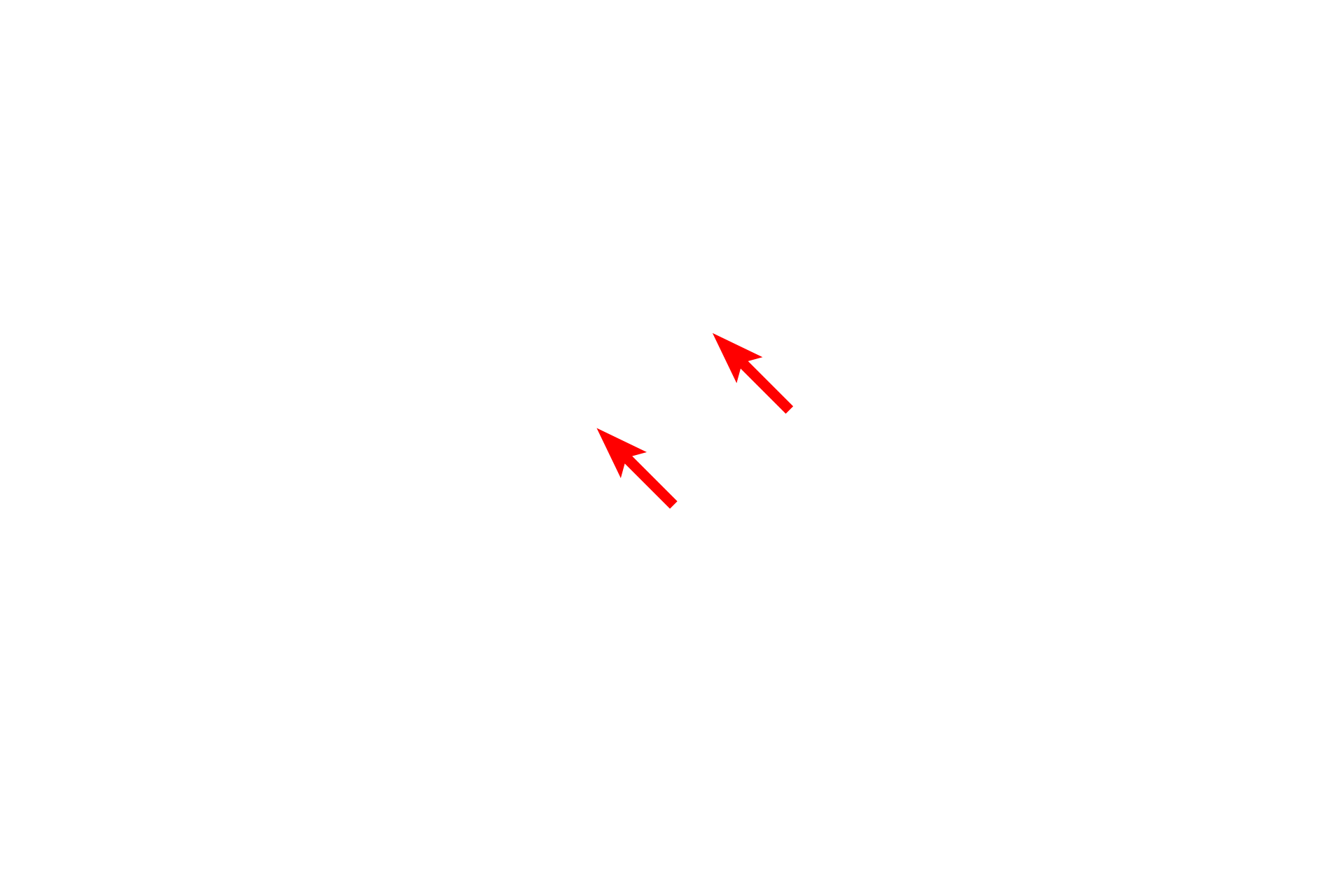
Pericytes, are multipotential cells with contractile properties that surround capillaries and postcapillary venules. They have highly branched cytoplasmic processes and are enclosed by a basal lamina that is continuous with that of the endothelial cells.

Basal laminae >
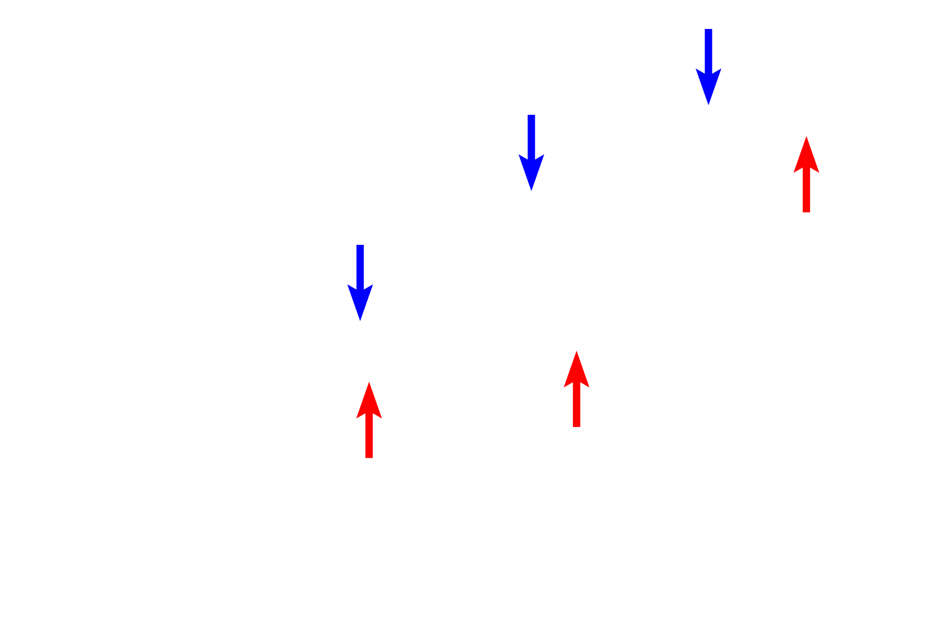
Both the endothelial cell and the gland cell are epithelial and thus each has a basal lamina. The red arrows indicate the basal lamina of the endocrine gland cell, the blue arrows indicate the basal lamina of the endothelium.