Continuous capillary
This electron micrograph shows a high magnification view of the basal surface of an endothelial cell. In a continuous capillary, no pores are present in the endothelium, and macromolecules are transported in both directions through the endothelial cytoplasm via pinocytotic vesicles. This bidirectional process is called transcytosis. Some molecules are able to diffuse across the endothelium, such as small ions, amino acids and gases. The endothelium rests on a basal lamina that it has produced. 45,000x

Endothelial cell
This electron micrograph shows a high magnification view of the basal surface of an endothelial cell. In a continuous capillary, no pores are present in the endothelium, and macromolecules are transported in both directions through the endothelial cytoplasm via pinocytotic vesicles. This bidirectional process is called transcytosis. Some molecules are able to diffuse across the endothelium, such as small ions, amino acids and gases. The endothelium rests on a basal lamina that it has produced. 45,000x

- Endothelial cell nucleus
This electron micrograph shows a high magnification view of the basal surface of an endothelial cell. In a continuous capillary, no pores are present in the endothelium, and macromolecules are transported in both directions through the endothelial cytoplasm via pinocytotic vesicles. This bidirectional process is called transcytosis. Some molecules are able to diffuse across the endothelium, such as small ions, amino acids and gases. The endothelium rests on a basal lamina that it has produced. 45,000x

- Basal lamina
This electron micrograph shows a high magnification view of the basal surface of an endothelial cell. In a continuous capillary, no pores are present in the endothelium, and macromolecules are transported in both directions through the endothelial cytoplasm via pinocytotic vesicles. This bidirectional process is called transcytosis. Some molecules are able to diffuse across the endothelium, such as small ions, amino acids and gases. The endothelium rests on a basal lamina that it has produced. 45,000x

- Pinocytic vesicles
This electron micrograph shows a high magnification view of the basal surface of an endothelial cell. In a continuous capillary, no pores are present in the endothelium, and macromolecules are transported in both directions through the endothelial cytoplasm via pinocytotic vesicles. This bidirectional process is called transcytosis. Some molecules are able to diffuse across the endothelium, such as small ions, amino acids and gases. The endothelium rests on a basal lamina that it has produced. 45,000x

Collagen fibrils >
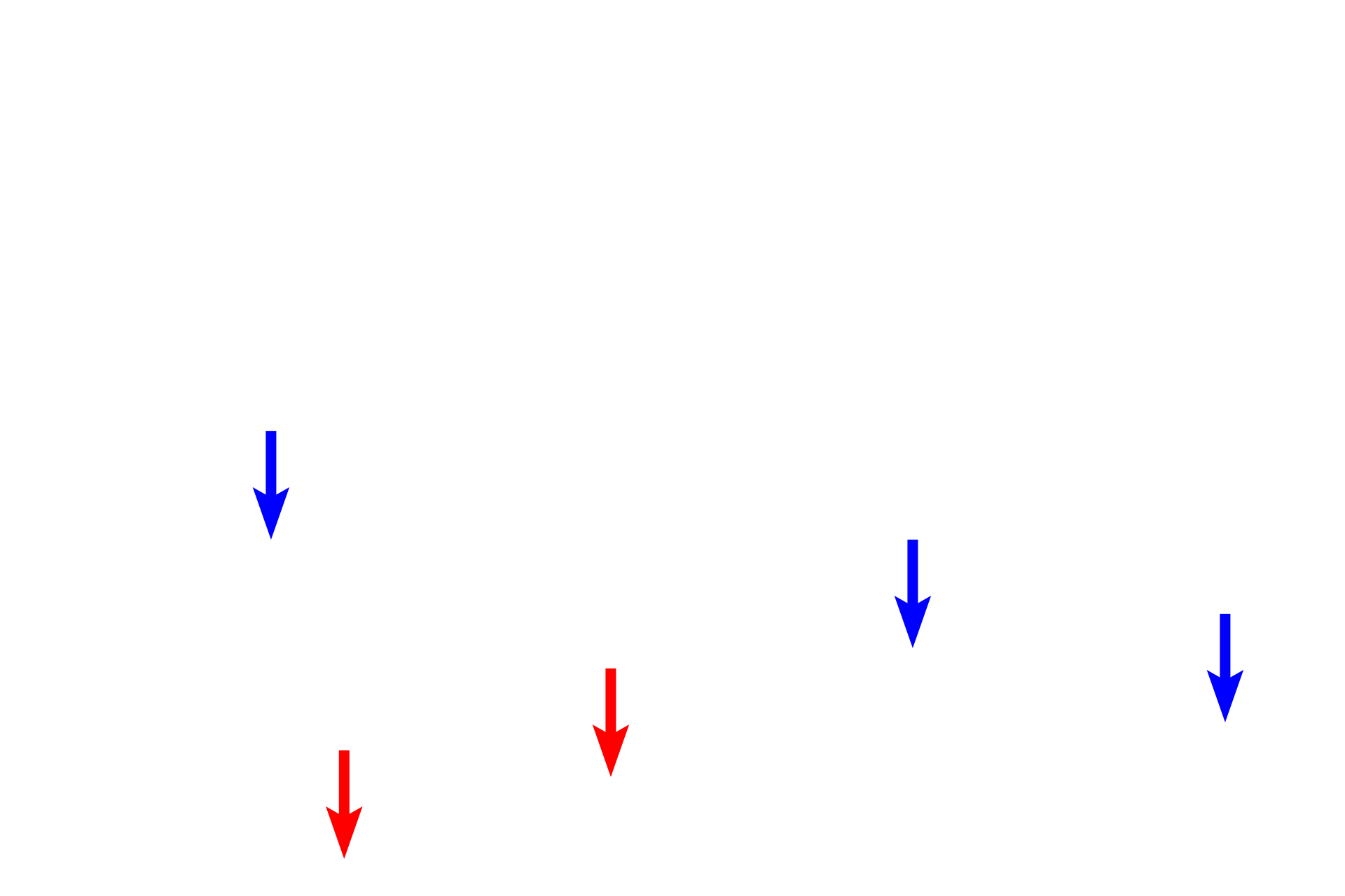
The very fine collagen fibrils immediately beneath the basal lamina (blue arrows) comprise the reticular lamina of the basement membrane and are composed of Type III collagen. The larger collagen fibrils (red arrows) are composed of Type I collagen.