
Anaphase
Anaphase begins abruptly as sister chromatids separate and are drawn toward opposite poles of the cell by the pushing action of the polar microtubules. This movement is also facilitated by the shortening of the kinetochore tubules. The end of anaphase is marked by the segregation of an identical set of chromosomes at each spindle pole.

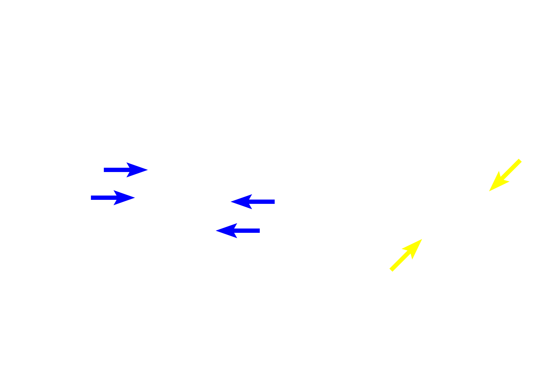
Early anaphase cell
Anaphase begins abruptly as sister chromatids separate and are drawn toward opposite poles of the cell by the pushing action of the polar microtubules. This movement is also facilitated by the shortening of the kinetochore tubules. The end of anaphase is marked by the segregation of an identical set of chromosomes at each spindle pole.

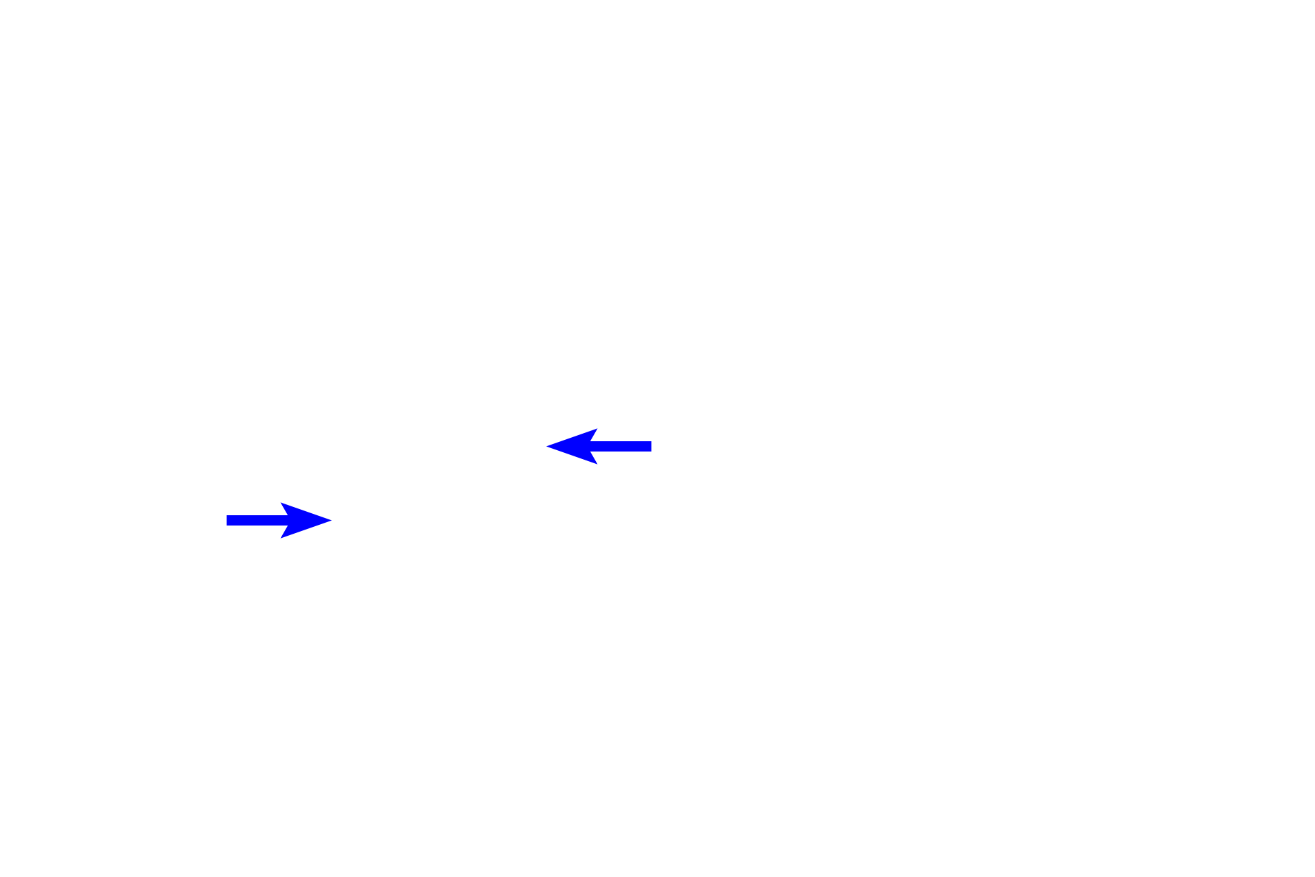
Mid-anaphase cell
Anaphase begins abruptly as sister chromatids separate and are drawn toward opposite poles of the cell by the pushing action of the polar microtubules. This movement is also facilitated by the shortening of the kinetochore tubules. The end of anaphase is marked by the segregation of an identical set of chromosomes at each spindle pole.
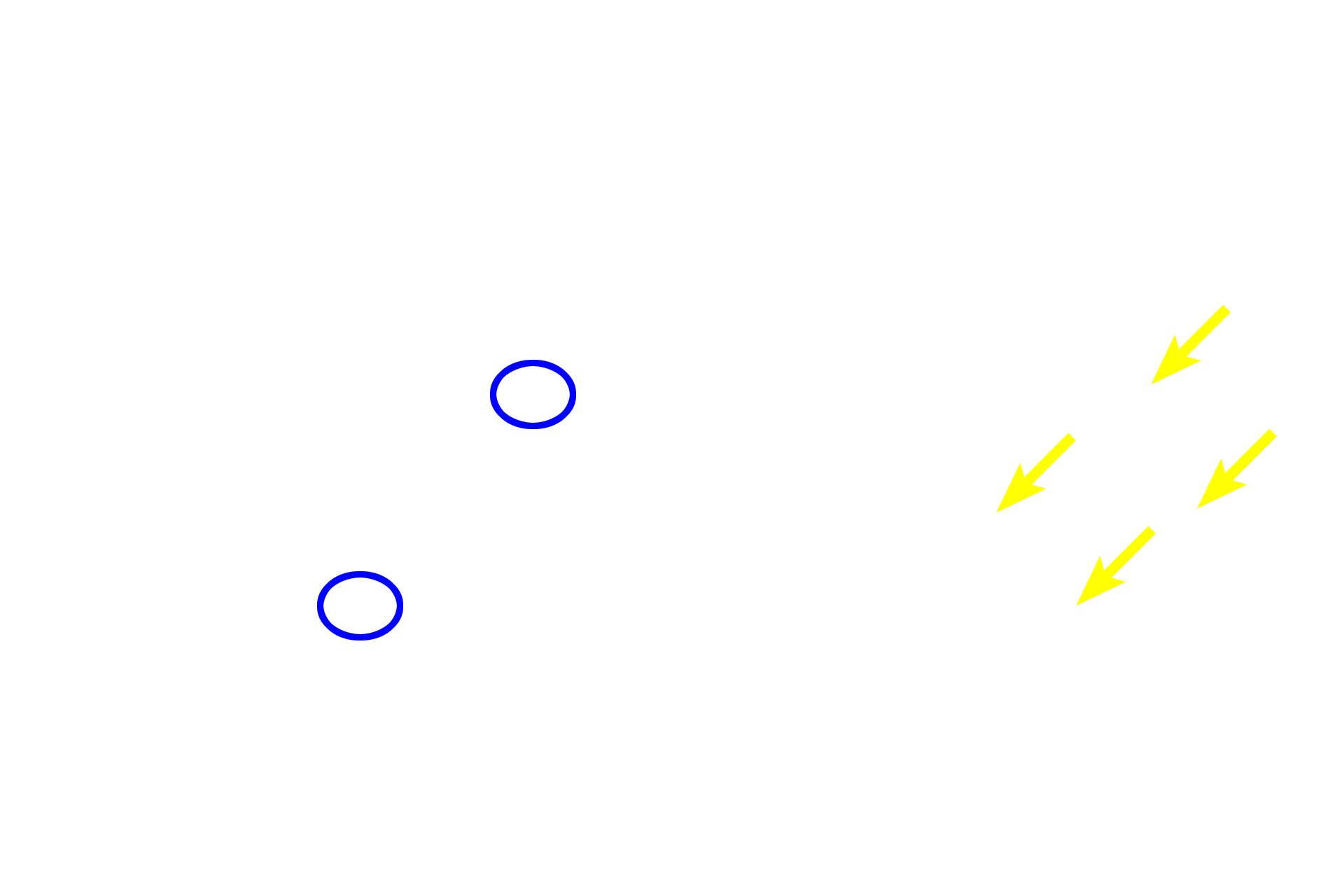
Chromosomes >
Sister chromatids are separated from each other following the breakdown of cohesive proteins holding them together at the centromere. Chromatids are then drawn to opposite poles by the pushing force of the polar microtubules and the pulling force of the kinetochore microtubules. Once separated, chromatids are referred to as chromosomes.
Centromeres
Sister chromatids are separated from each other following the breakdown of cohesive proteins holding them together at the centromere. Chromatids are then drawn to opposite poles by the pushing force of the polar microtubules and the pulling force of the kinetochore microtubules. Once separated, chromatids are referred to as chromosomes.
Centrosomes
Sister chromatids are separated from each other following the breakdown of cohesive proteins holding them together at the centromere. Chromatids are then drawn to opposite poles by the pushing force of the polar microtubules and the pulling force of the kinetochore microtubules. Once separated, chromatids are referred to as chromosomes.
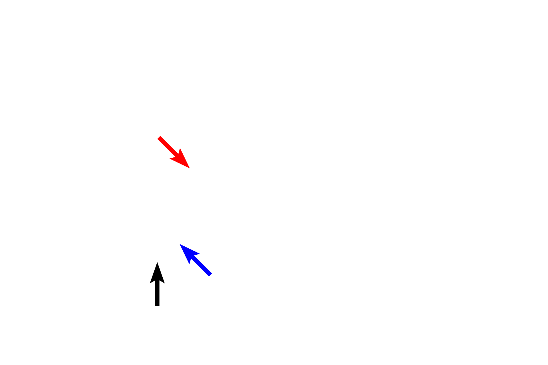
Mitotic spindle >
The mitotic spindle consists of three types of microtubules. Kinetochore microtubules (blue arrow) attach to chromatids, drawing them toward the spindle pole. Polar microtubules (red arrow) overlap in the center of the spindle, interacting with each other to push the spindle poles apart. Astral microtubules (black arrow) extend away from the spindle to provide anchorage for the spindle.