
Globe: Iris
This image shows two sections through the iris which is part of the middle or uveal tunic and is continuous with the ciliary body. The inner margin of the iris forms the sphincter bordering the pupillary opening. The iris is located just anterior to the lens and regulates the amount of light that enters the eye. 100x

Iris >
The iris is a donut-shaped structure surrounding the pupil. The anterior surface of the iris is irregular and rough, covered by a discontinuous layer of cells. Most of the core of the iris consists of loose connective tissue with many melanocytes and blood vessels. Contraction of smooth muscle groups in the iris alters the diameter of the pupil and, therefore, alters the amount of light entering the eye.
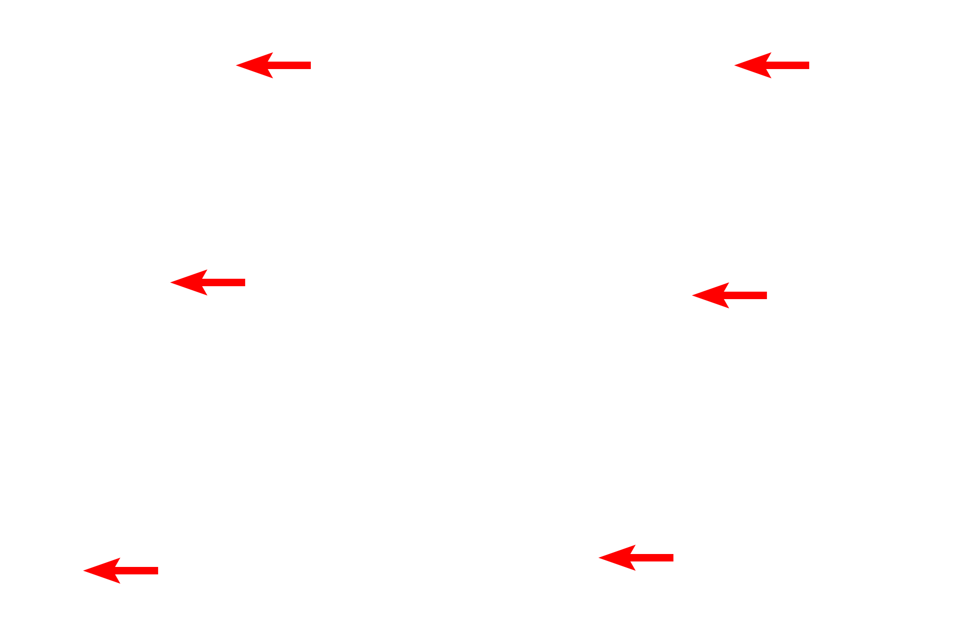
- Iris epithelium >
The epithelium covering the posterior surface is a continuation of the non-sensory retina and consists of two layers: a heavily pigmented epithelium next to the posterior chamber and a less pigmented layer of myoepithelial cells from which radially arranged processes extend into the stroma, forming dilator pupillae muscle. Contraction of this muscle results in an increase in pupillary diameter.
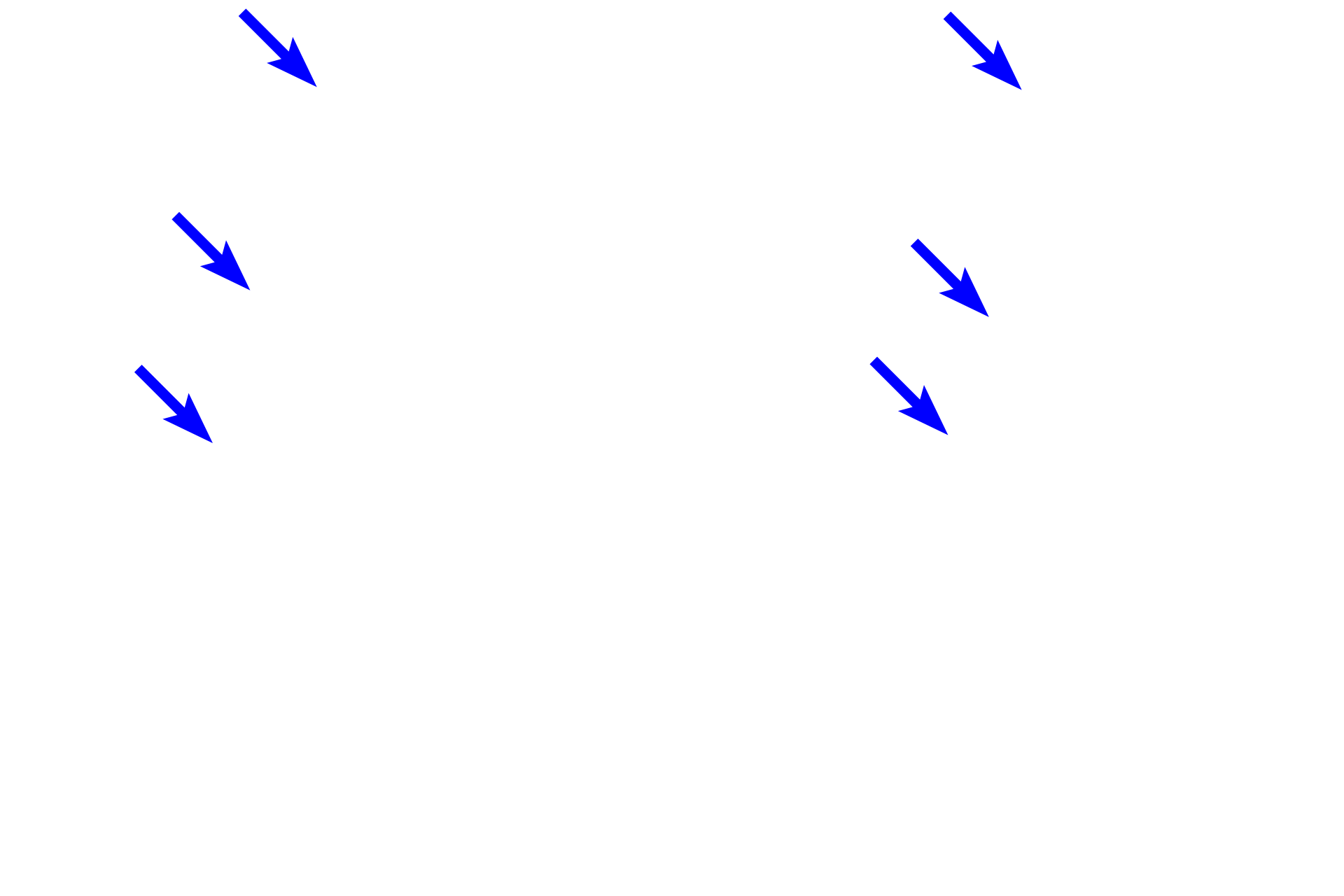
- Dilator pupillae muscle
The epithelium covering the posterior surface is a continuation of the non-sensory retina and consists of two layers: a heavily pigmented epithelium next to the posterior chamber and a less pigmented layer of myoepithelial cells from which radially arranged processes extend into the stroma, forming dilator pupillae muscle. Contraction of this muscle results in an increase in pupillary diameter.

- Constrictor pupillae > muscle
The constrictor pupillae muscle decreases the diameter of the pupil and is the antagonistic muscle of the dilator pupillae. The constrictor pupillae muscle consists of smooth muscle fibers located at the internal perimeter of the iris. These muscle fibers are oriented circumferentially around the pupillary rim of the iris.
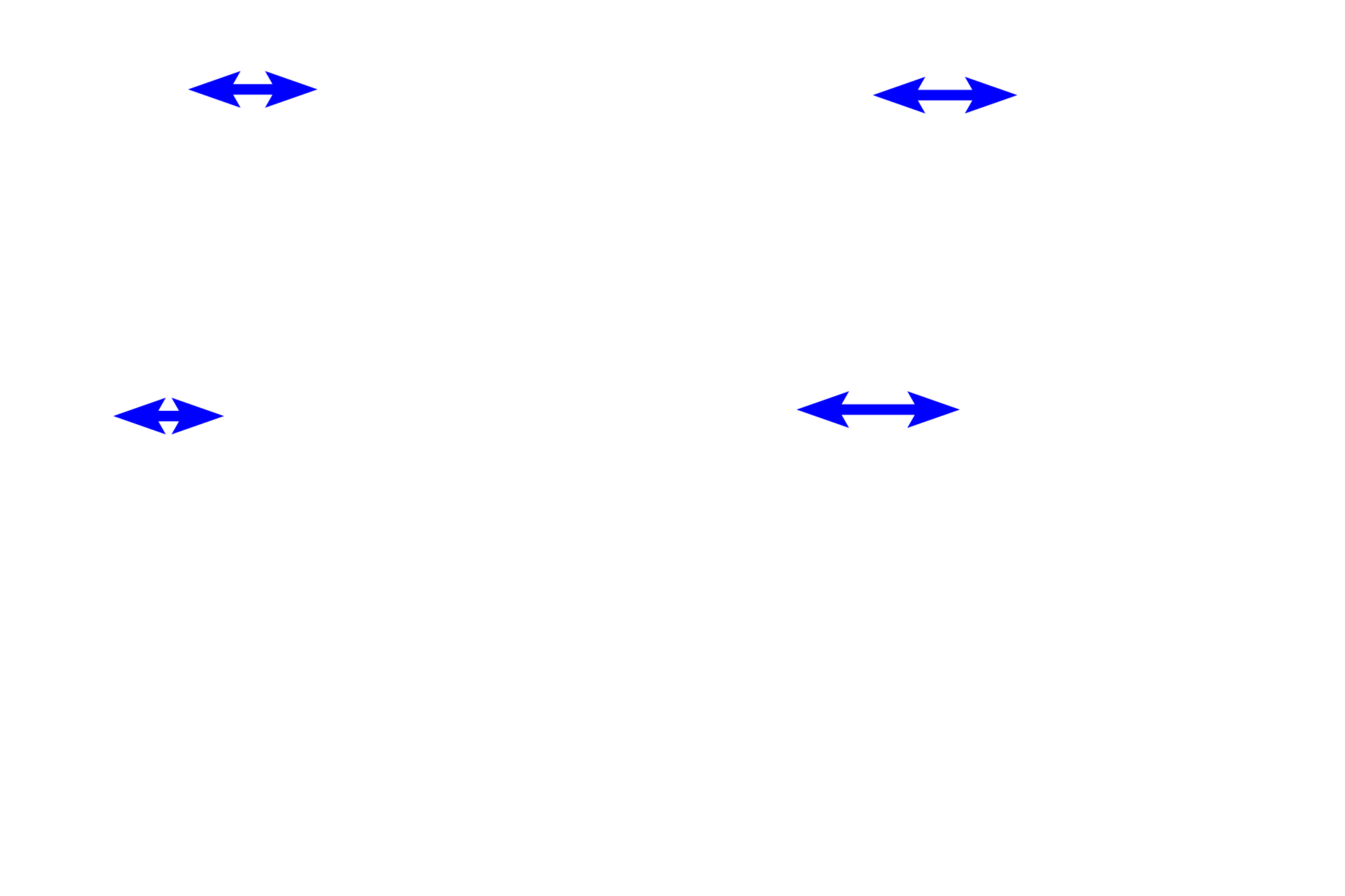
- Stroma >
Stroma, formimg the bulk of the iris, is composed of loose connective tissue, containing fibroblasts and melanocytes. The stroma is highly vascular. The anterior surface of the iris has numerous grooves and ridges and is not covered by an epithelium.

Lens >
The bulk of the lens is made primarily of elongated lens fibers that are modified cells of the lens epithelium. The lens epithelium is present only on the anterior and equatorial regions of the lens and consists of a single layer of cuboidal cells. The outermost layer of the lens is a capsule, which represents the highly thickened basal lamina of the lens epithelium that lies inside the capsule.
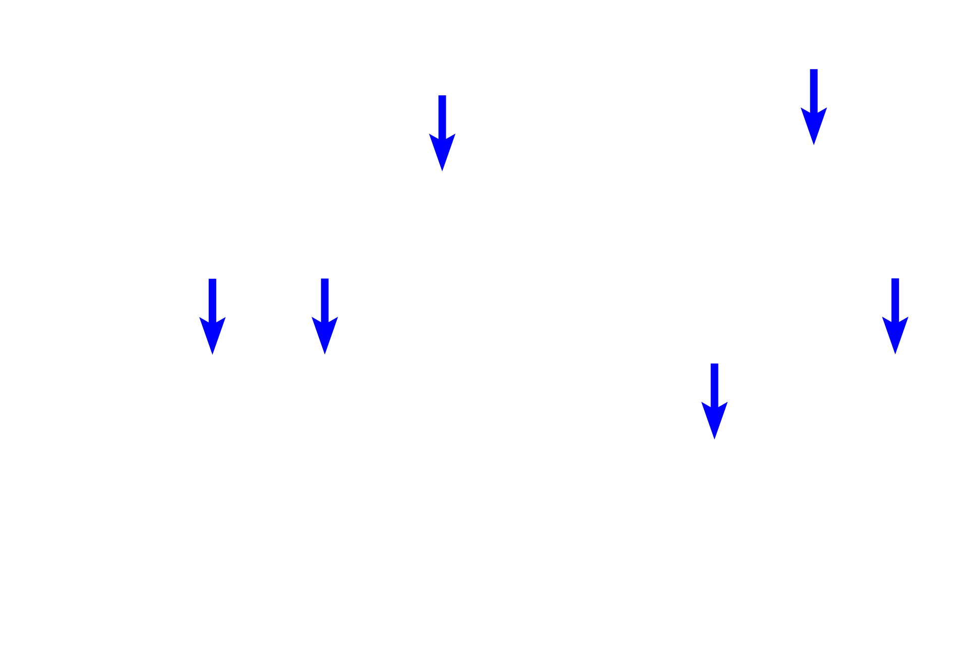
- Lens fibers
The bulk of the lens is made primarily of elongated lens fibers that are modified cells of the lens epithelium. The lens epithelium is present only on the anterior and equatorial regions of the lens and consists of a single layer of cuboidal cells. The outermost layer of the lens is a capsule, which represents the highly thickened basal lamina of the lens epithelium that lies inside the capsule.
- Lens epithelium
The bulk of the lens is made primarily of elongated lens fibers that are modified cells of the lens epithelium. The lens epithelium is present only on the anterior and equatorial regions of the lens and consists of a single layer of cuboidal cells. The outermost layer of the lens is a capsule, which represents the highly thickened basal lamina of the lens epithelium that lies inside the capsule.
- Lens capsule
The bulk of the lens is made primarily of elongated lens fibers that are modified cells of the lens epithelium. The lens epithelium is present only on the anterior and equatorial regions of the lens and consists of a single layer of cuboidal cells. The outermost layer of the lens is a capsule, which represents the highly thickened basal lamina of the lens epithelium that lies inside the capsule.
Anterior chamber >
The anterior chamber lies anterior to the iris and the lens.
Image source >
Image taken of a slide from the University of Iowa collection. Inset: Arizona Veterinary Diagnostic Laboratory
 PREVIOUS