Main Menu » Tissues » Muscle » Skeletal » Skeletal 17
This is blank.

Skeletal muscle

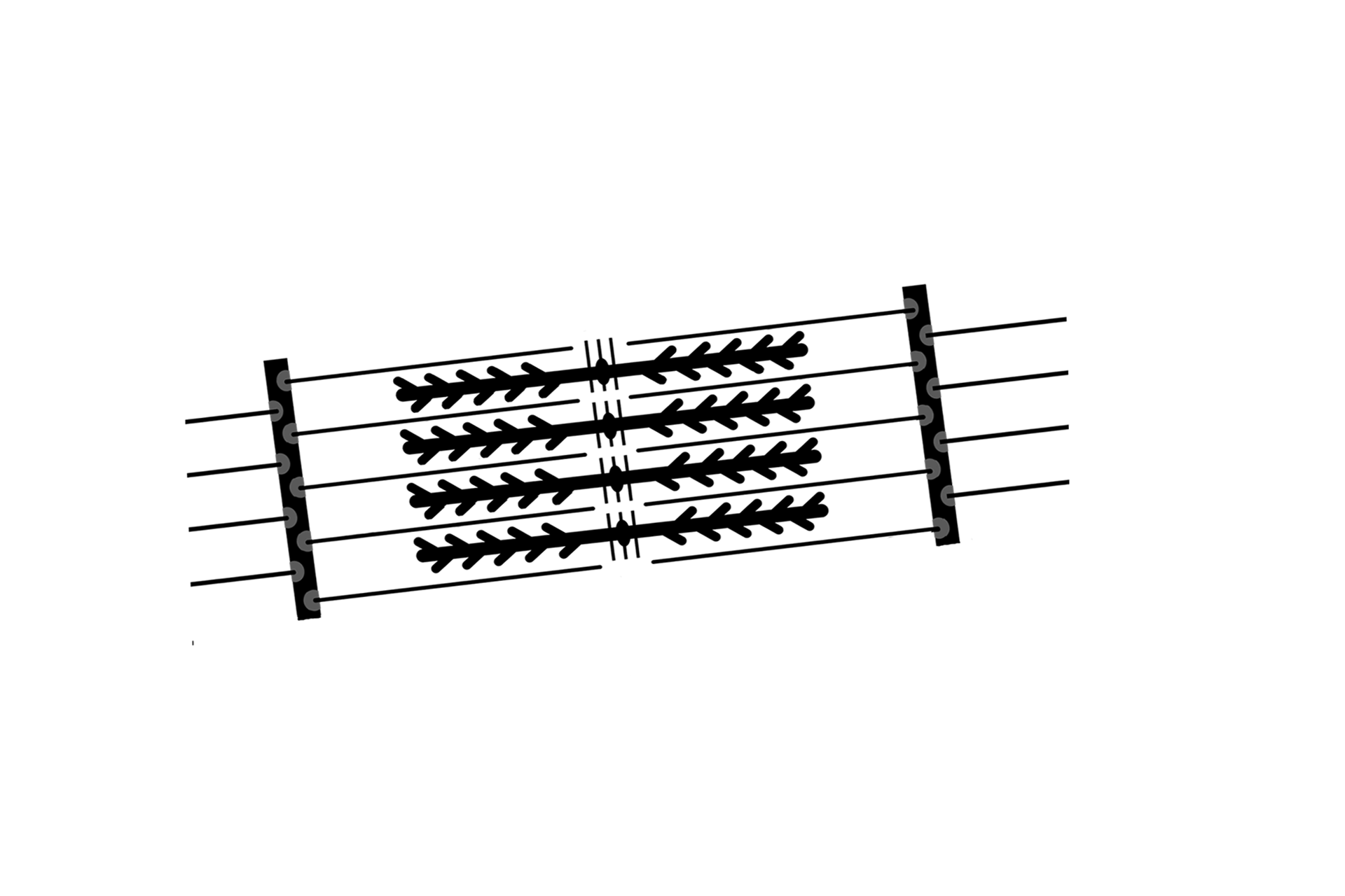
This higher magnification electron micrograph of skeletal muscle shows greater detail of the myofibrils and overlapping myofilaments. 60,000x

Myofibrils > <p>Myofibrils contain the contractile thin and thick myofilaments. The overlap of the myofilaments is the basis for the sliding filament mechanism of contraction. The pattern of overlap remains in register across the width of the myofibril, which produces the characteristic banding seen in striated muscle.</p>

Myofibrils >

Myofibrils contain the contractile thin and thick myofilaments. The overlap of the myofilaments is the basis for the sliding filament mechanism of contraction. The pattern of overlap remains in register across the width of the myofibril, which produces the characteristic banding seen in striated muscle.

Sarcomeres > <p>The sarcomere is the contractile unit of striated muscle and extends from one Z line to an adjacent Z line. During contraction, the width of the sarcomere narrows. The width of the A band remains constant, but both the I and H bands narrow as the areas of overlap of thin and thick myofilaments increase.</p>

Sarcomeres >

The sarcomere is the contractile unit of striated muscle and extends from one Z line to an adjacent Z line. During contraction, the width of the sarcomere narrows. The width of the A band remains constant, but both the I and H bands narrow as the areas of overlap of thin and thick myofilaments increase.

A band <p>The sarcomere is the contractile unit of striated muscle and extends from one Z line to an adjacent Z line. During contraction, the width of the sarcomere narrows. The width of the A band remains constant, but both the I and H bands narrow as the areas of overlap of thin and thick myofilaments increase.</p>

A band

The sarcomere is the contractile unit of striated muscle and extends from one Z line to an adjacent Z line. During contraction, the width of the sarcomere narrows. The width of the A band remains constant, but both the I and H bands narrow as the areas of overlap of thin and thick myofilaments increase.

 - H band <p>The sarcomere is the contractile unit of striated muscle and extends from one Z line to an adjacent Z line. During contraction, the width of the sarcomere narrows. The width of the A band remains constant, but both the I and H bands narrow as the areas of overlap of thin and thick myofilaments increase.</p>

- H band

The sarcomere is the contractile unit of striated muscle and extends from one Z line to an adjacent Z line. During contraction, the width of the sarcomere narrows. The width of the A band remains constant, but both the I and H bands narrow as the areas of overlap of thin and thick myofilaments increase.

 - M line <p>The sarcomere is the contractile unit of striated muscle and extends from one Z line to an adjacent Z line. During contraction, the width of the sarcomere narrows. The width of the A band remains constant, but both the I and H bands narrow as the areas of overlap of thin and thick myofilaments increase.</p>

- M line

The sarcomere is the contractile unit of striated muscle and extends from one Z line to an adjacent Z line. During contraction, the width of the sarcomere narrows. The width of the A band remains constant, but both the I and H bands narrow as the areas of overlap of thin and thick myofilaments increase.

I band <p>The sarcomere is the contractile unit of striated muscle and extends from one Z line to an adjacent Z line. During contraction, the width of the sarcomere narrows. The width of the A band remains constant, but both the I and H bands narrow as the areas of overlap of thin and thick myofilaments increase.</p>

I band

The sarcomere is the contractile unit of striated muscle and extends from one Z line to an adjacent Z line. During contraction, the width of the sarcomere narrows. The width of the A band remains constant, but both the I and H bands narrow as the areas of overlap of thin and thick myofilaments increase.

 - Z lines <p>The sarcomere is the contractile unit of striated muscle and extends from one Z line to an adjacent Z line. During contraction, the width of the sarcomere narrows. The width of the A band remains constant, but both the I and H bands narrow as the areas of overlap of thin and thick myofilaments increase.</p>

- Z lines

The sarcomere is the contractile unit of striated muscle and extends from one Z line to an adjacent Z line. During contraction, the width of the sarcomere narrows. The width of the A band remains constant, but both the I and H bands narrow as the areas of overlap of thin and thick myofilaments increase.

Thin myofilaments > <p>Thin myofilaments consist primarily of polymerized globular actin molecules. Globular actin polymerizes, forming the filament which has binding sites for myosin of the thick myofilament. Tropomyosin and troponin proteins spiral around the filament, regulating the interaction of myosin with the thin filaments. The thin myofilament is anchored to the Z line, consisting of alpha-actinin and Cap Z proteins.</p>

Thin myofilaments >

Thin myofilaments consist primarily of polymerized globular actin molecules. Globular actin polymerizes, forming the filament which has binding sites for myosin of the thick myofilament. Tropomyosin and troponin proteins spiral around the filament, regulating the interaction of myosin with the thin filaments. The thin myofilament is anchored to the Z line, consisting of alpha-actinin and Cap Z proteins.

Thick myofilaments > <p>Thick myofilaments are composed of myosin II, consisting of two heavy chains and four light chains. Head groups on the myosin bind to the actin of the thin myofilaments. The conformational changes of the head groups move the thin myofilament relative to the thick myofilament, a process referred to as the sliding filament mechanism.</p>

Thick myofilaments >

Thick myofilaments are composed of myosin II, consisting of two heavy chains and four light chains. Head groups on the myosin bind to the actin of the thin myofilaments. The conformational changes of the head groups move the thin myofilament relative to the thick myofilament, a process referred to as the sliding filament mechanism.

Sarcomere overlay > <p>Diagrammatic representation of the major structural elements of the sarcomere.</p>

Sarcomere overlay >

Diagrammatic representation of the major structural elements of the sarcomere.

Triad > <p>The triad is a structural feature of skeletal muscle that is responsible for the regulation of excitation-contraction coupling. It is formed by the close apposition of the T-tubule and expanded cisterns of the sarcoplasmic reticulum. Each skeletal muscle fiber has many thousands of triads which in skeletal muscle are located at the level of the junction of the A and I bands.</p>

Triad >

The triad is a structural feature of skeletal muscle that is responsible for the regulation of excitation-contraction coupling. It is formed by the close apposition of the T-tubule and expanded cisterns of the sarcoplasmic reticulum. Each skeletal muscle fiber has many thousands of triads which in skeletal muscle are located at the level of the junction of the A and I bands.

 - T tubules > <p>The T tubule is an invagination of the sarcolemma and serves to carry the membrane depolarization into the depths of the cell. This rapid transmission into the center of the cell insures a coordinated contraction across its width.</p>

- T tubules >

The T tubule is an invagination of the sarcolemma and serves to carry the membrane depolarization into the depths of the cell. This rapid transmission into the center of the cell insures a coordinated contraction across its width.

 - Sarcoplasmic reticulum > <p>The smooth endoplasmic in muscle fibers is called the sarcoplasmic reticulum. Two cisterns of the sarcoplasmic reticulum lie immediately adjacent the T tubule, thereby forming the triad. These cisterns serve as calcium stores. The depolarization carried by the T tubules causes the release of this calcium into the cytoplasm. Increased intracellular calcium is the signal that initiates muscle contraction. The sarcoplasmic reticulum also takes up calcium after contraction.</p>

- Sarcoplasmic reticulum >

The smooth endoplasmic in muscle fibers is called the sarcoplasmic reticulum. Two cisterns of the sarcoplasmic reticulum lie immediately adjacent the T tubule, thereby forming the triad. These cisterns serve as calcium stores. The depolarization carried by the T tubules causes the release of this calcium into the cytoplasm. Increased intracellular calcium is the signal that initiates muscle contraction. The sarcoplasmic reticulum also takes up calcium after contraction.