
Skeletal muscle
The banding pattern seen in the high magnification light micrograph (left) is compared with that seen by electron microscopy (right). The bands in all the individual myofibrils are closely aligned, resulting in the overall banding pattern seen by light microscopy. Sparse cytoplasm (sarcoplasm) between the myofibrils contains mitochondria, sarcoplasmic reticulum and glycogen. 1,000x, 15,000x
Myofibrils >
Myofibrils almost totally fill the cytoplasm of skeletal muscle fibers and contain the contractile thick and thin myofilaments. The myofilament overlap remains in register across each myofibril, thus accounting for the banding pattern within each myofibril. Alignment of all myofibrils in the cell produces the banding visible at the light microscopic level. Due to their dense packing within the fibers, individual myofibrils are not visible in the light micrograph.
Myofilament organization >
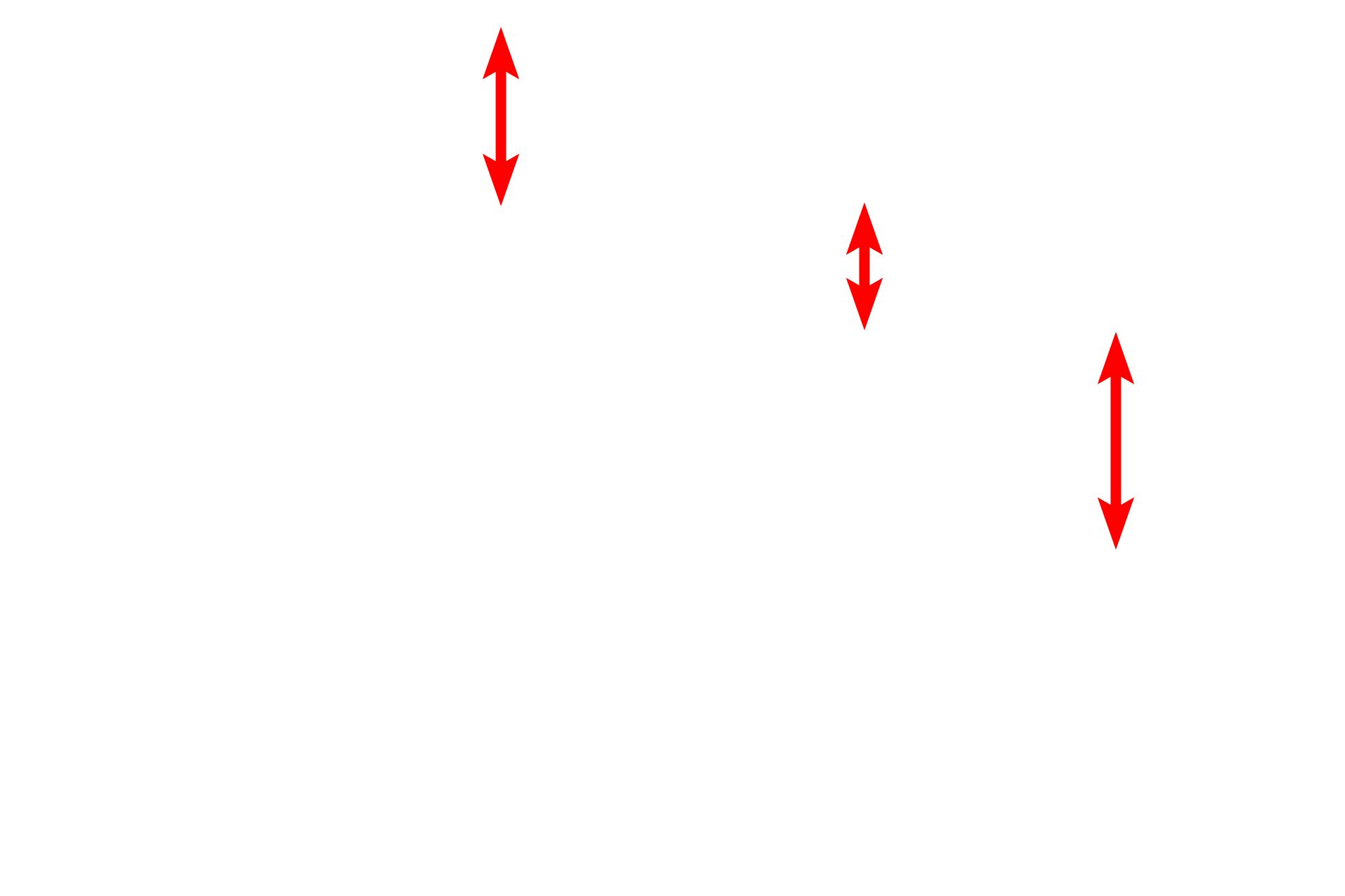
This diagram illustrates a single sarcomere and the myofilaments that compose it. Note how the thick and thin myofilaments overlap, producing the striations visible by both light and electron microscopy. This molecular organization of the sarcomere will be correlated with the image seen by electron microscopy.
Sarcomeres >
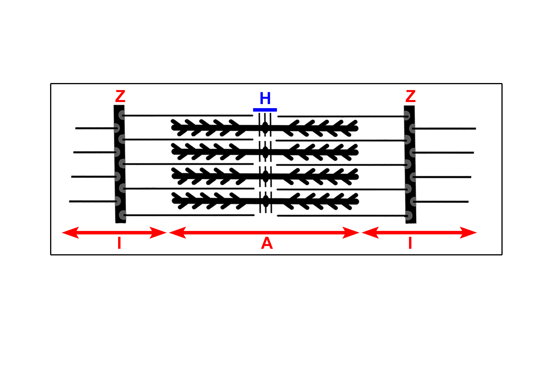
The sarcomere is the contractile unit of striated muscles and extends from one Z line to an adjacent Z line. During contraction, the interval between adjacent Z lines narrows, i.e., the sarcomere shortens. The width of the A band remains constant during contraction, but both the I and H bands shorten as the areas of overlap of thin and thick myofilaments increases.
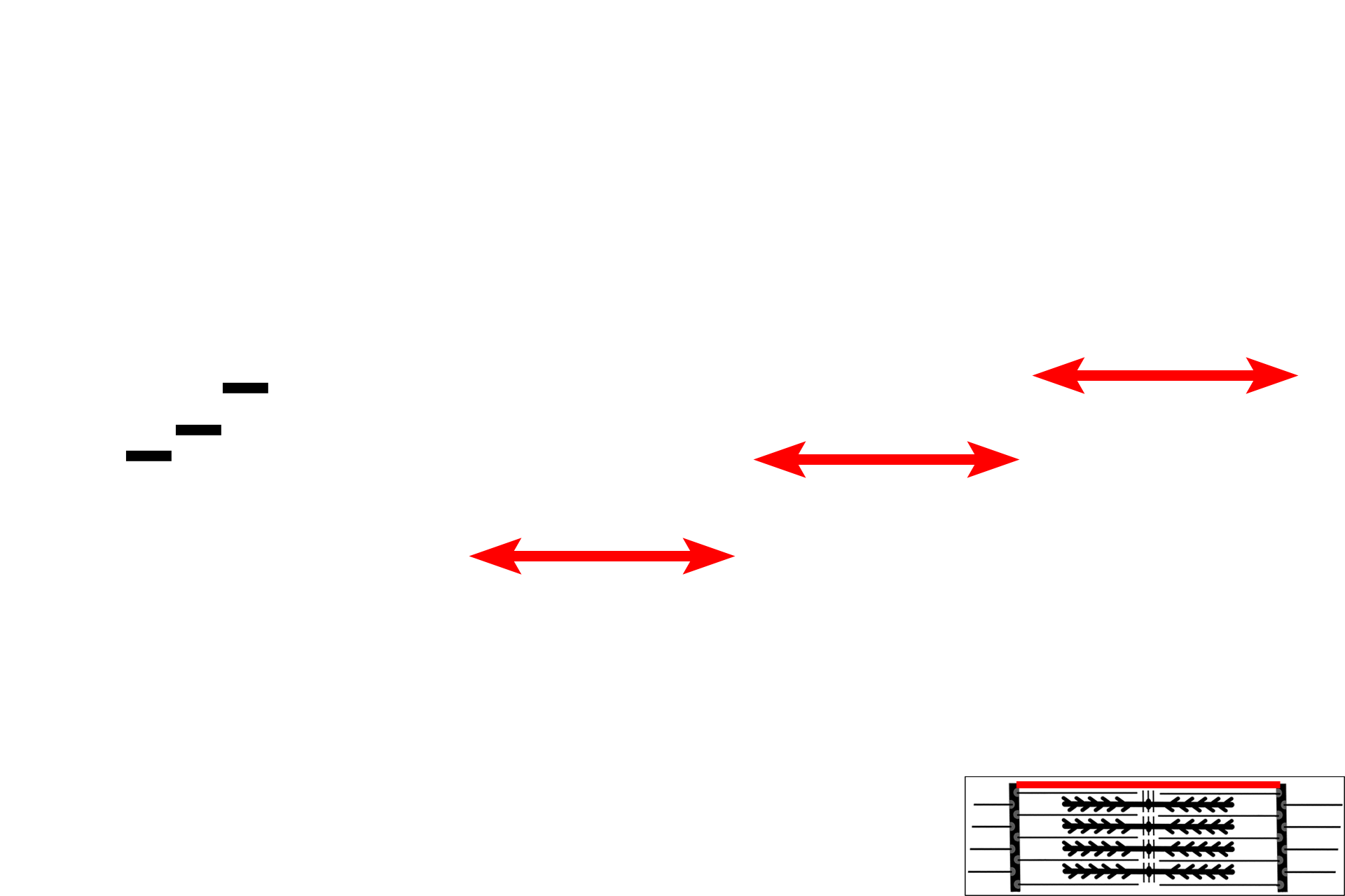
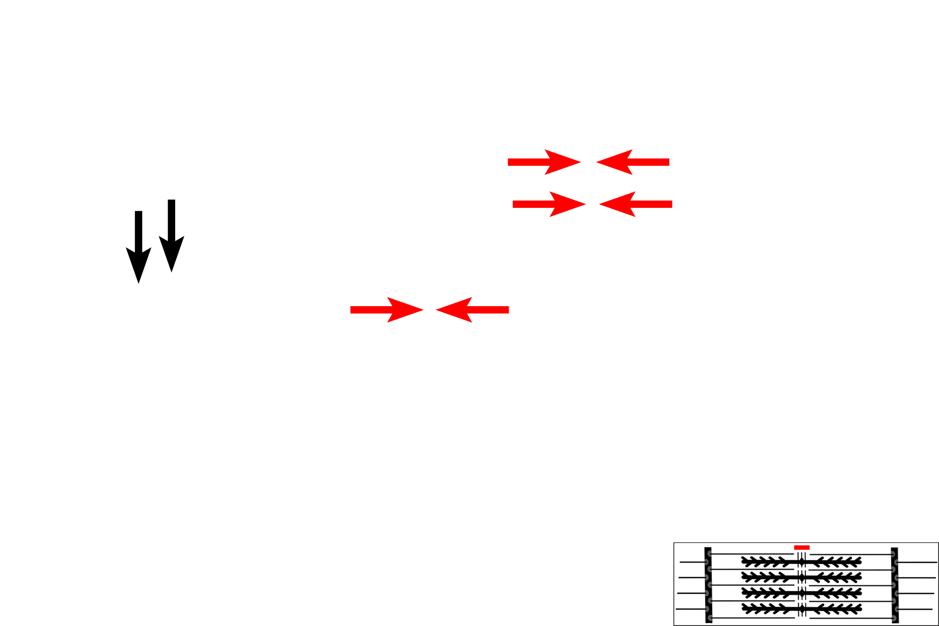
A band >
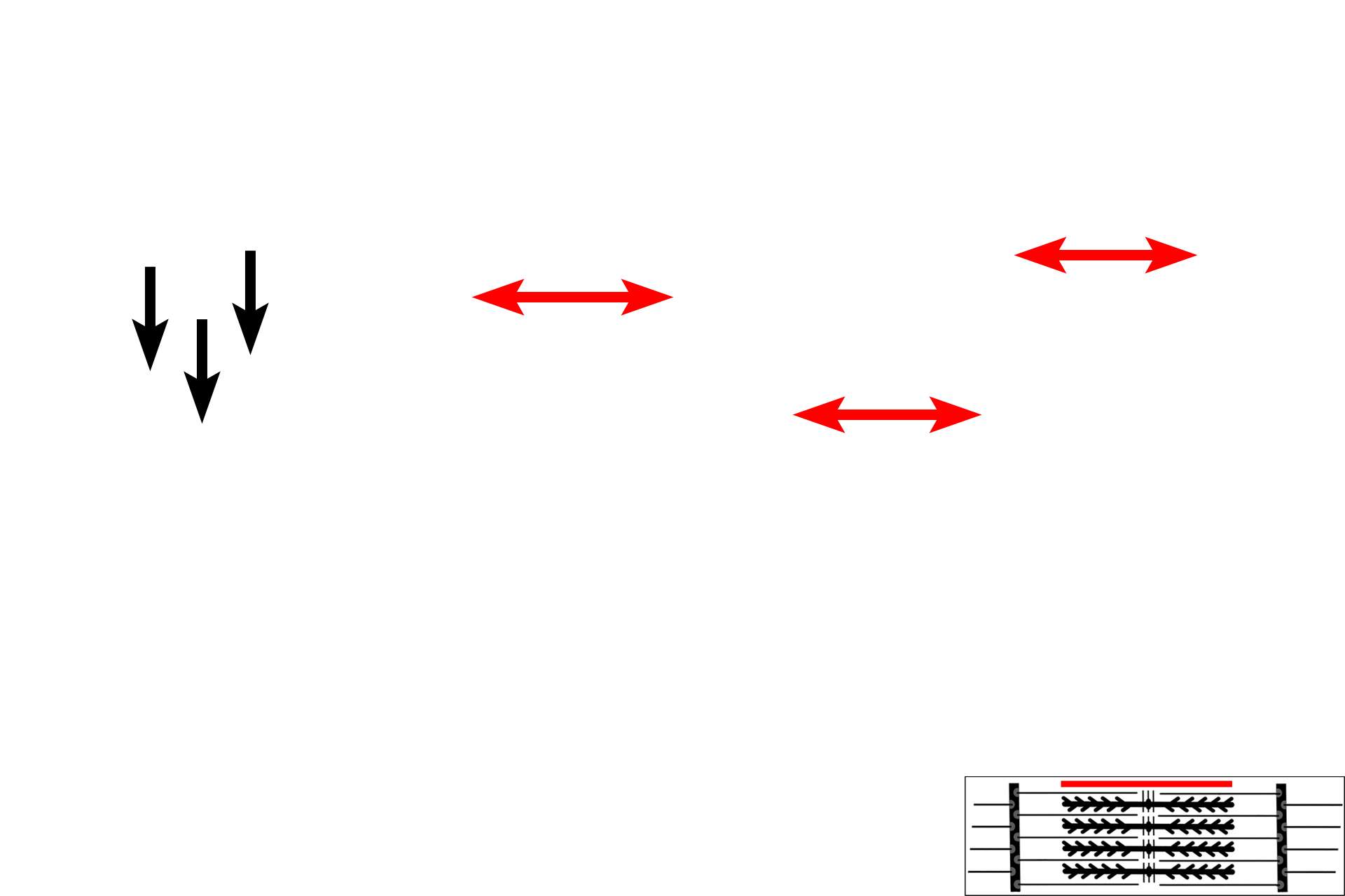
The A band corresponds to the length of the thick myofilaments. Thin myofilaments overlap with thick filaments at the margins of the A band, but they do not extend all the way to its center, leaving a lighter central region. This thin filament-free central region of the A band is the H band, with a centermost portion called the M line. During contraction, the width of the A band remains constant.
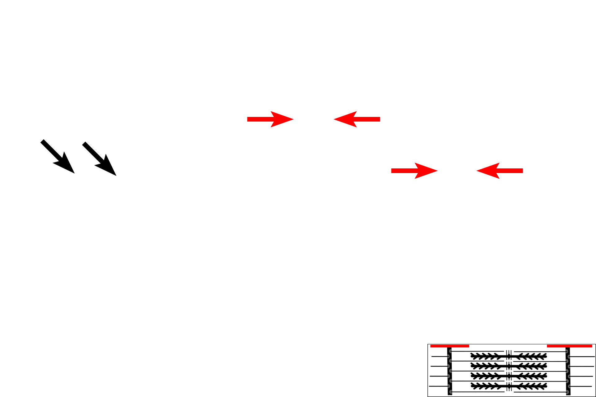
- H band >
The H band is the central region of the A band which lacks thin filaments. This region of the A band decreases in width as the ends of the thin filaments are drawn toward the center of the sarcomere during contraction.
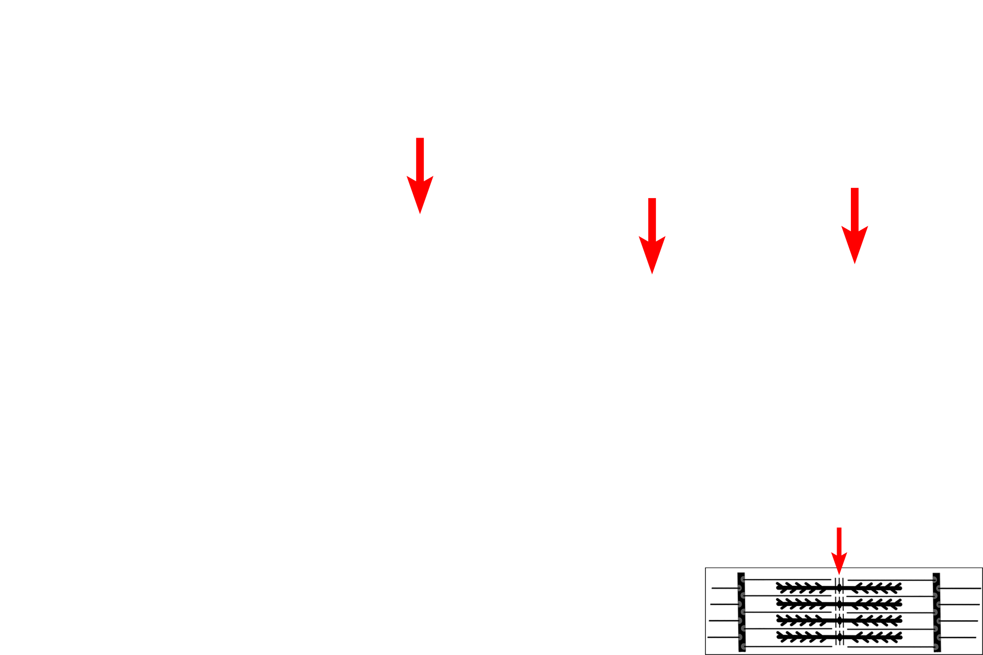
- M line >
The M line bisects the H band and contains myosin-binding proteins that maintain the alignment of the thick myofilaments.
I band >
The I band, or light band, contains only thin myofilaments and is bisected by the Z line. During contraction, the width of A band decreases.
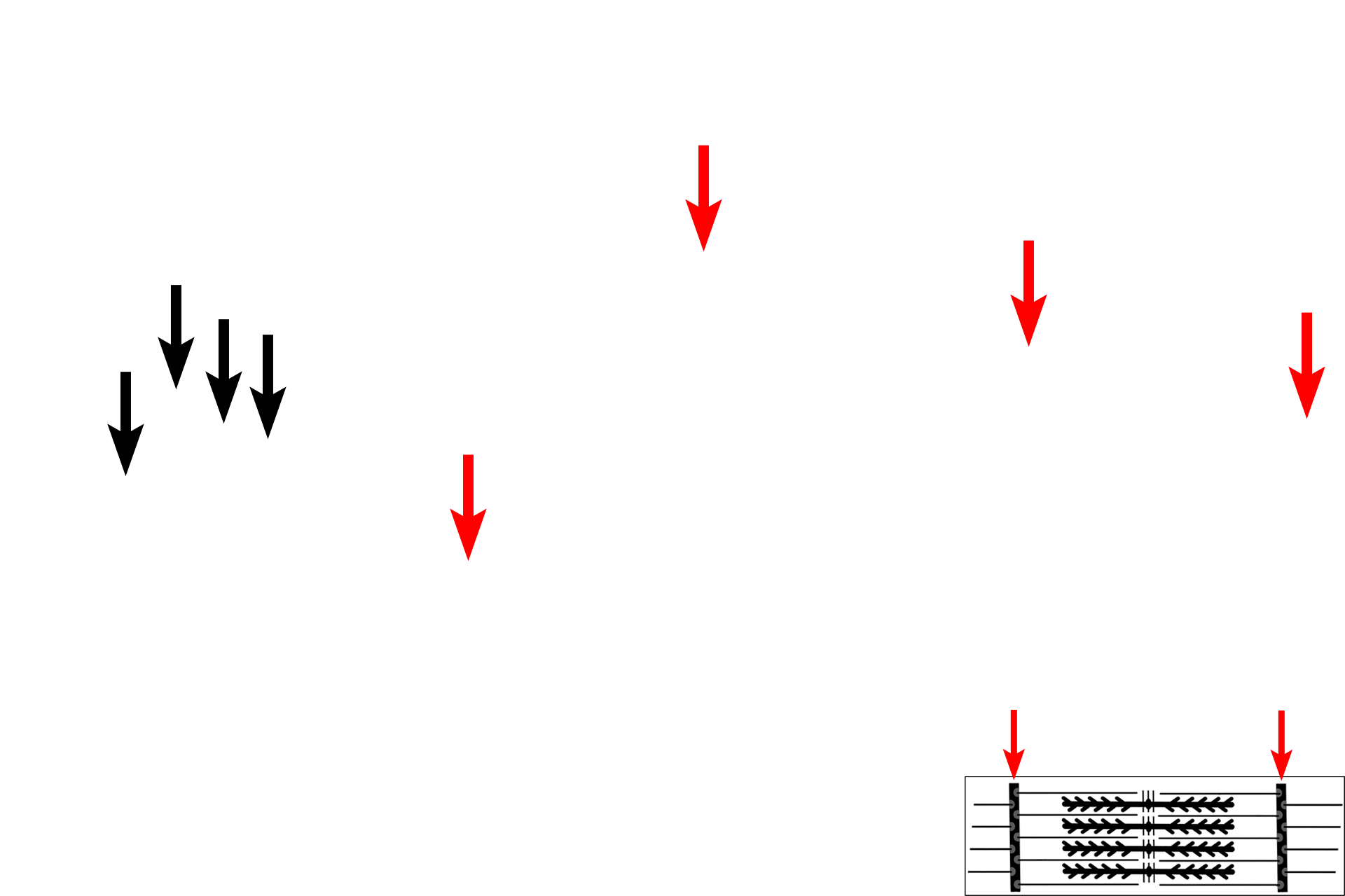
- Z line >
The Z line lies in the center of the I band and is the attachment site for thin myofilaments. The Z line is composed primarily of the protein alpha-actinin. The region from one Z line to the next is called a sarcomere.
Mitochondria
The banding pattern seen in the high magnification light micrograph (left) is compared with that seen by electron microscopy (right). The bands in all the individual myofibrils are closely aligned, resulting in the overall banding pattern seen by light microscopy. Sparse cytoplasm (sarcoplasm) between the myofibrils contains mitochondria, sarcoplasmic reticulum and glycogen. 1,000x, 15,000x

Sarcoplasmic reticulum (SER)
The banding pattern seen in the high magnification light micrograph (left) is compared with that seen by electron microscopy (right). The bands in all the individual myofibrils are closely aligned, resulting in the overall banding pattern seen by light microscopy. Sparse cytoplasm (sarcoplasm) between the myofibrils contains mitochondria, sarcoplasmic reticulum and glycogen. 1,000x, 15,000x

Glycogen
The banding pattern seen in the high magnification light micrograph (left) is compared with that seen by electron microscopy (right). The bands in all the individual myofibrils are closely aligned, resulting in the overall banding pattern seen by light microscopy. Sparse cytoplasm (sarcoplasm) between the myofibrils contains mitochondria, sarcoplasmic reticulum and glycogen. 1,000x, 15,000x