
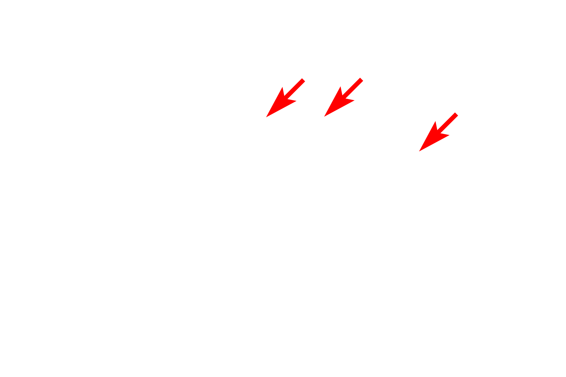
Golgi apparatus
The Golgi, usually located near the nucleus, consists of flattened, membranous sacs. These sacs receive newly synthesized proteins from the RER via transport vesicles. The vesicles fuse with the forming face of the Golgi, and their proteins are post-translationally modified, e.g., glycosylated or phosphorylated, and packaged on the maturing face of the Golgi for transport through the cell.

RER
The Golgi, usually located near the nucleus, consists of flattened, membranous sacs. These sacs receive newly synthesized proteins from the RER via transport vesicles. The vesicles fuse with the forming face of the Golgi, and their proteins are post-translationally modified, e.g., glycosylated or phosphorylated, and packaged on the maturing face of the Golgi for transport through the cell.
Transport vesicles
The Golgi, usually located near the nucleus, consists of flattened, membranous sacs. These sacs receive newly synthesized proteins from the RER via transport vesicles. The vesicles fuse with the forming face of the Golgi, and their proteins are post-translationally modified, e.g., glycosylated or phosphorylated, and packaged on the maturing face of the Golgi for transport through the cell.

Golgi apparatus
The Golgi, usually located near the nucleus, consists of flattened, membranous sacs. These sacs receive newly synthesized proteins from the RER via transport vesicles. The vesicles fuse with the forming face of the Golgi, and their proteins are post-translationally modified, e.g., glycosylated or phosphorylated, and packaged on the maturing face of the Golgi for transport through the cell.

- Forming (cis) face
The Golgi, usually located near the nucleus, consists of flattened, membranous sacs. These sacs receive newly synthesized proteins from the RER via transport vesicles. The vesicles fuse with the forming face of the Golgi, and their proteins are post-translationally modified, e.g., glycosylated or phosphorylated, and packaged on the maturing face of the Golgi for transport through the cell.

- Maturing (trans) face
The Golgi, usually located near the nucleus, consists of flattened, membranous sacs. These sacs receive newly synthesized proteins from the RER via transport vesicles. The vesicles fuse with the forming face of the Golgi, and their proteins are post-translationally modified, e.g., glycosylated or phosphorylated, and packaged on the maturing face of the Golgi for transport through the cell.
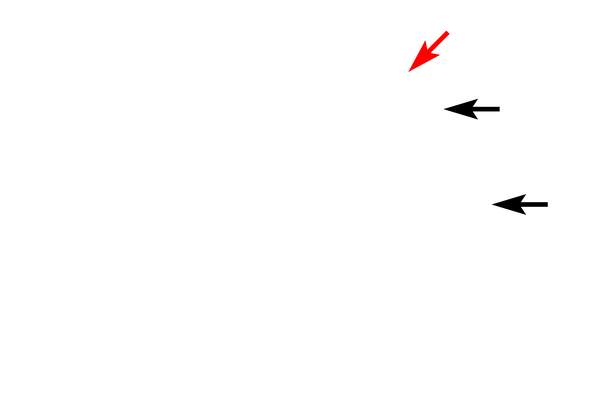
Secretory granules >
Secretory granules (black arrows), also referred to as secretory vesicles, are one of several types of vesicles derived from the Golgi. Secretory granules contain proteins synthesized in the RER, that are secreted from the cells by exocytosis (red arrow). Examples of proteins transported by secretory granules include digestive enzymes, peptide hormones, and extracellular matrix components.
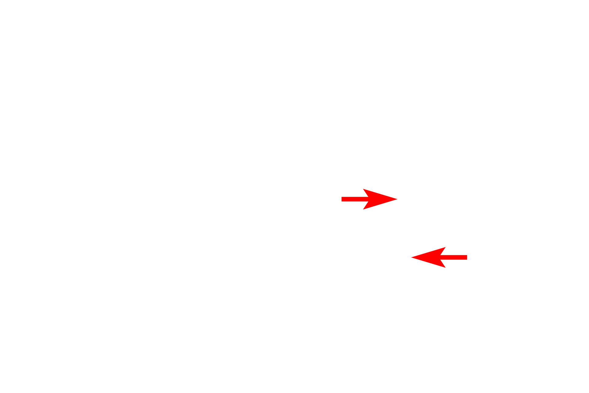
Pre-lysosomal vesicles >
The Golgi also produces pre-lysosomal vesicles containing hydrolytic enzymes. These vesicles form primary lysosomes by fusion with endosomes, containing materials derived by endocytosis, phagocytosis or by autophagocytosis of worn-out organelles. The hydrolytic enzymes in primary lysosomes break down the endosomal contents, thus transitioning into secondary lysosomes.