
Golgi apparatus
The location of the Golgi is clearly evident in this H&E stained section of the pituitary gland. The location of the Golgi apparatus appears as a pale-staining region of the cytoplasm adjacent to the nucleus, surrounded by the secretory granules it produces. The large Golgi displaces the granules thus resulting is a lightly stained region, referred to as “negative Golgi” image. The structure of the Golgi apparatus cannot be seen with H&E staining. 1000x
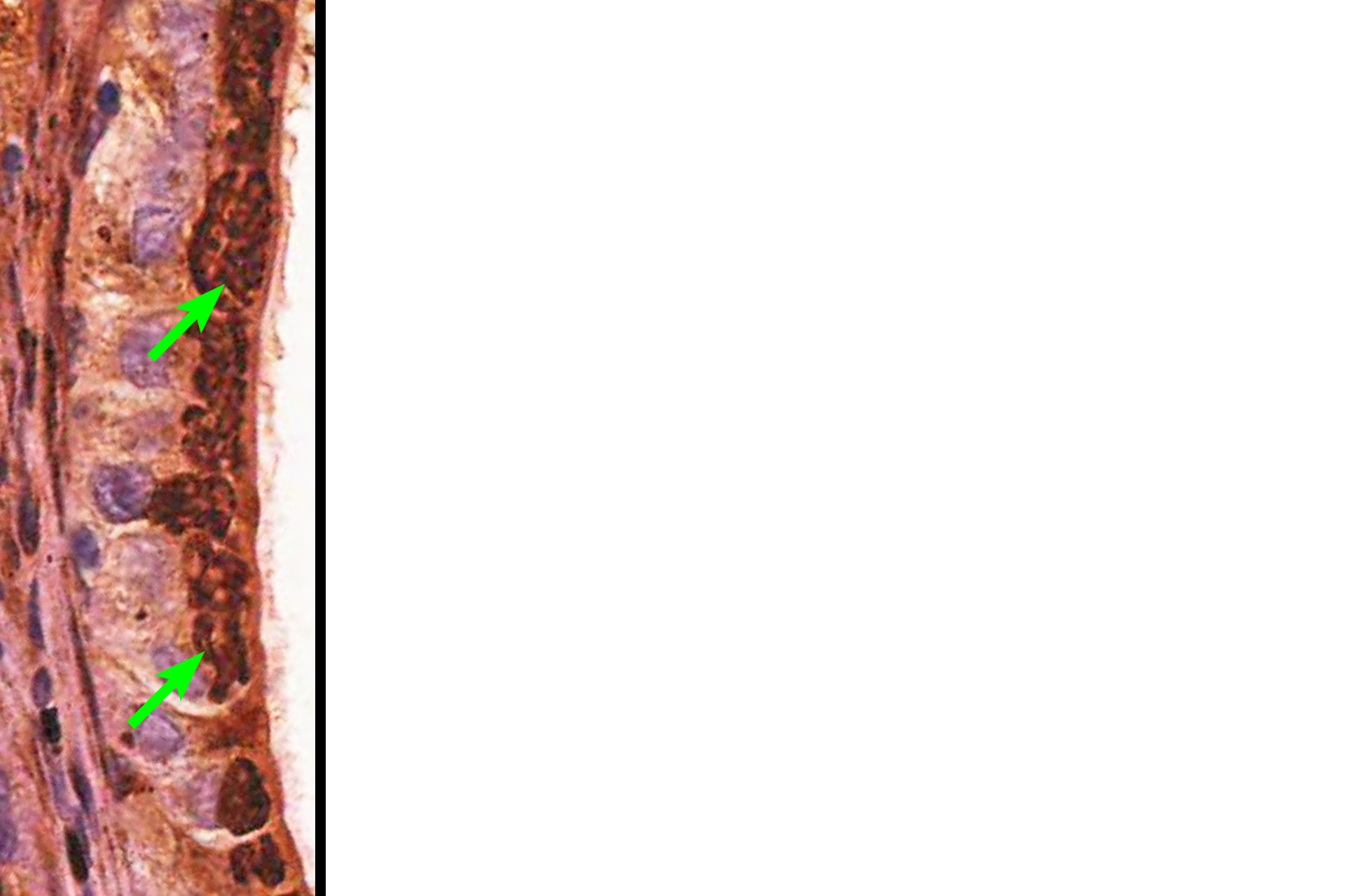
Golgi apparatus (silver) >
This image shows a row of epithelium cells in which the Golgi apparatus has been stained by the silver method which does label the Golgi membranes. Here the Golgi can been seen as a threadlike network in the apical region of the cells. Conventional staining methods, like H&E, do not reveal these membranes.
Secretory granules
The location of the Golgi is clearly evident in this H&E stained section of the pituitary gland. The location of the Golgi apparatus appears as a pale-staining region of the cytoplasm adjacent to the nucleus, surrounded by the secretory granules it produces. The large Golgi displaces the granules thus resulting is a lightly stained region, referred to as “negative Golgi” image. The structure of the Golgi apparatus cannot be seen with H&E staining. 1000x
Nuclei
The location of the Golgi is clearly evident in this H&E stained section of the pituitary gland. The location of the Golgi apparatus appears as a pale-staining region of the cytoplasm adjacent to the nucleus, surrounded by the secretory granules it produces. The large Golgi displaces the granules thus resulting is a lightly stained region, referred to as “negative Golgi” image. The structure of the Golgi apparatus cannot be seen with H&E staining. 1000x
- Nucleoli
The location of the Golgi is clearly evident in this H&E stained section of the pituitary gland. The location of the Golgi apparatus appears as a pale-staining region of the cytoplasm adjacent to the nucleus, surrounded by the secretory granules it produces. The large Golgi displaces the granules thus resulting is a lightly stained region, referred to as “negative Golgi” image. The structure of the Golgi apparatus cannot be seen with H&E staining. 1000x