
Placenta: 1st trimester
The placenta is a union of maternal and fetal tissues. The decidua, the maternal portion, is derived from decidual cells of the uterine endometrium. The fetal portion, the chorion, is an extraembryonic tissue. The placenta has two major functions: maintenance of pregnancy through the production of hormones; and exchange of nutrients and oxygen from the mother with wastes and carbon dioxide from the embryo/fetus, respectively. 4x
Embryo >
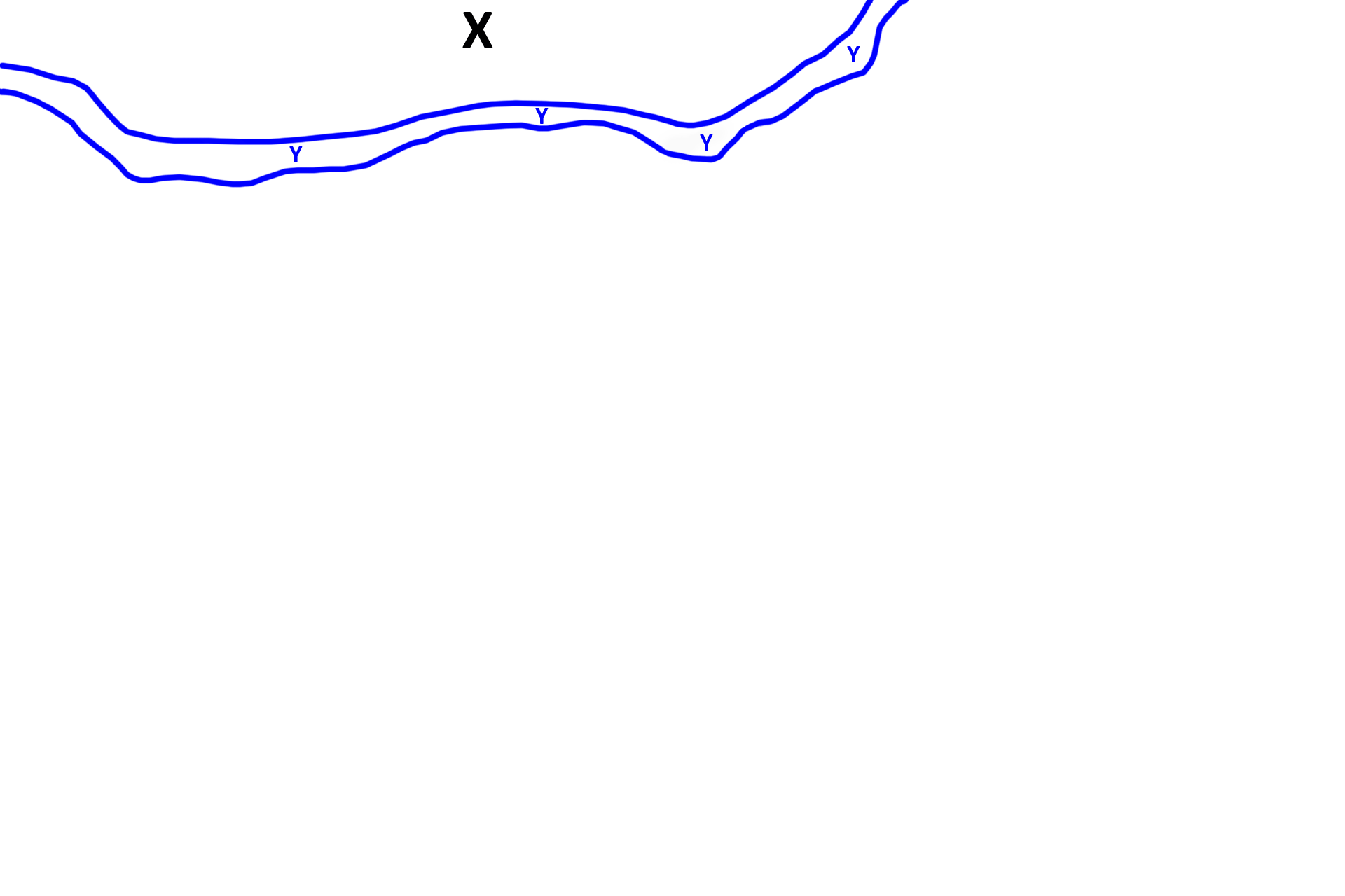
The embryo would be located at “X.” An extraembryonic membrane, the amnion, is also absent in this image; its location is indicated by Y and the blue lines. The amnion is a fluid-filled sac that surrounds the embryo/fetus, suspending and protecting it. The amnion becomes continuous with the chorion as pregnancy progresses.
Chorion >
Chorion, the embryonic portion of the placenta, surrounds the amnion (and, therefore, the embryo/fetus), forming the outer rind of the embryonic placenta. The chorion has three components. A chorionic plate lies closest to the embryo. Chorionic villi extend from the plate into an intervillous space filled with maternal blood. The third component, the (cyto)trophoblastic shell, forms an outer rind of the embryonic placenta and is fused with the maternal decidua.
- Chorionic plate >
The chorionic plate is composed of extraembryonic connective tissue covered by an epithelium, the trophoblast, which faces the intervillous space. The trophoblast is composed of two layers: cytotrophoblast lies adjacent to the connective tissue and syncytiotrophoblast faces the intervillous space. The syncytiotrophoblast is a multinucleated syncytium formed from the cytotrophoblast.
- Chorionic villi >
Chorionic villi are fingerlike projections of the chorion that form the bulk of the embryonic placenta. Chorionic villi extend from the chorionic plate into the intervillous space, which is filled with maternal blood supplied by the spiral arteries. Therefore, the villi are the site of exchange of nutrients and wastes in the placenta. Like the chorionic plate, villi have a central core of connective tissue covered by the double-layered trophoblast.
- Intervillous space
Chorionic villi are fingerlike projections of the chorion that form the bulk of the embryonic placenta. Chorionic villi extend from the chorionic plate into the intervillous space, which is filled with maternal blood supplied by the spiral arteries. Therefore, the villi are the site of exchange of nutrients and wastes in the placenta. Like the chorionic plate, villi have a central core of connective tissue covered by the double-layered trophoblast.
- Trophoblastic shell >
The (cyto)trophoblastic shell is a thickened layer of cytotrophoblastic cells that forms the outer perimeter of the embryonic placenta and lies adjacent to the maternal portion of the placenta, the decidua. The (cyto)trophoblastic shell is lined with syncytiotrophoblast on the surface facing the intervillous space.
Decidua >
The maternal portion of the placenta, the decidua, is composed of a layer of decidual cells. Decidual cells differentiate from stromal cells in the functional zone of the uterine endometrium a few days after implantation occurs. The decidua is separated from the trophoblastic shell by a fibrinoid layer.
Fibrinoid
The maternal portion of the placenta, the decidua, is composed of a layer of decidual cells. Decidual cells differentiate from stromal cells in the functional zone of the uterine endometrium a few days after implantation occurs. The decidua is separated from the trophoblastic shell by a fibrinoid layer.

Maternal blood vessels >
Maternal blood vessels are visible in the decidual layer. Uterine arteries supply the spiral arteries that spurt blood into the intervillous space. Blood drains from the intervillous space into veins that eventually form the uterine veins.
Next images
The next image shows a portion of the embryonic surface of the placenta on the left and the maternal surface on the right, similar to the areas outlined by the rectangles.