
Placenta: 1st trimester
The embryonic side of the placenta is seen in greater detail in the left image, while the maternal side is shown in the right image. 100x, 100x

Chorionic plate >
The amnion was removed in this preparation; it would have enclosed the amniotic cavity surrounding the embryo and would have been located at the upper border of this image. The chorionic plate (bars), surrounding the amnion, is composed of extraembryonic connective tissue covered by a double layer of trophoblastic cells, seen as a thin, purple line (arrows) on the lower edge of the plate.
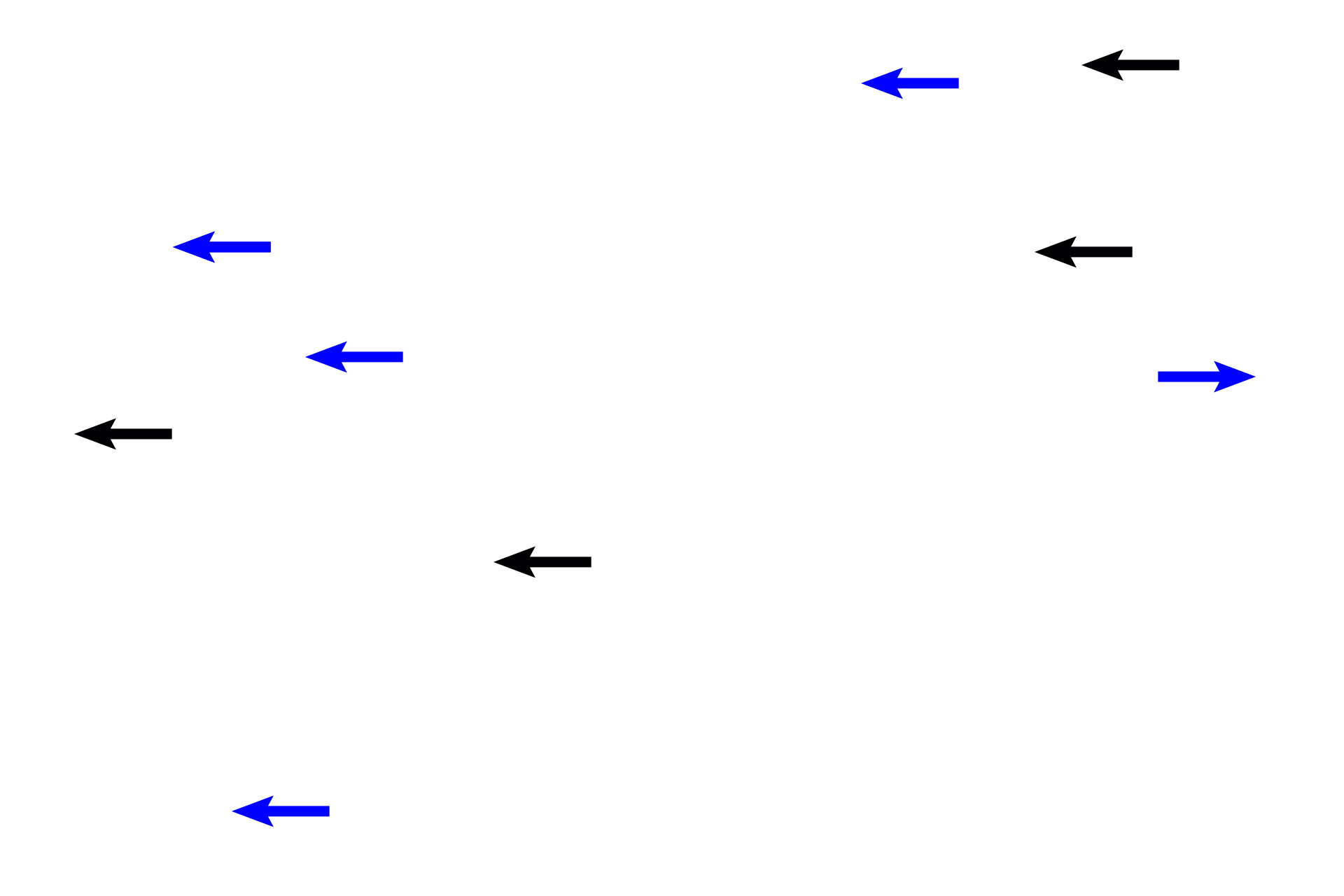
Chorionic villi >
Chorionic villi, the functional components of the embryonic placenta, are composed of a central core of embryonic connective tissue (blue arrows) covered by a double layer of trophoblastic epithelium (black arrows). Chorionic villi extend from the chorionic plate and branch from this trunk like roots of a tree. Villi contain embryonic blood vessels involved with exchange of nutrients and wastes with maternal blood.

- Stem villus >
Stem villi originate from the chorionic plate. Numerous, branching villi, termed floating villi, extend from each stem villus into the intervillous space, where they are bathed by maternal blood.
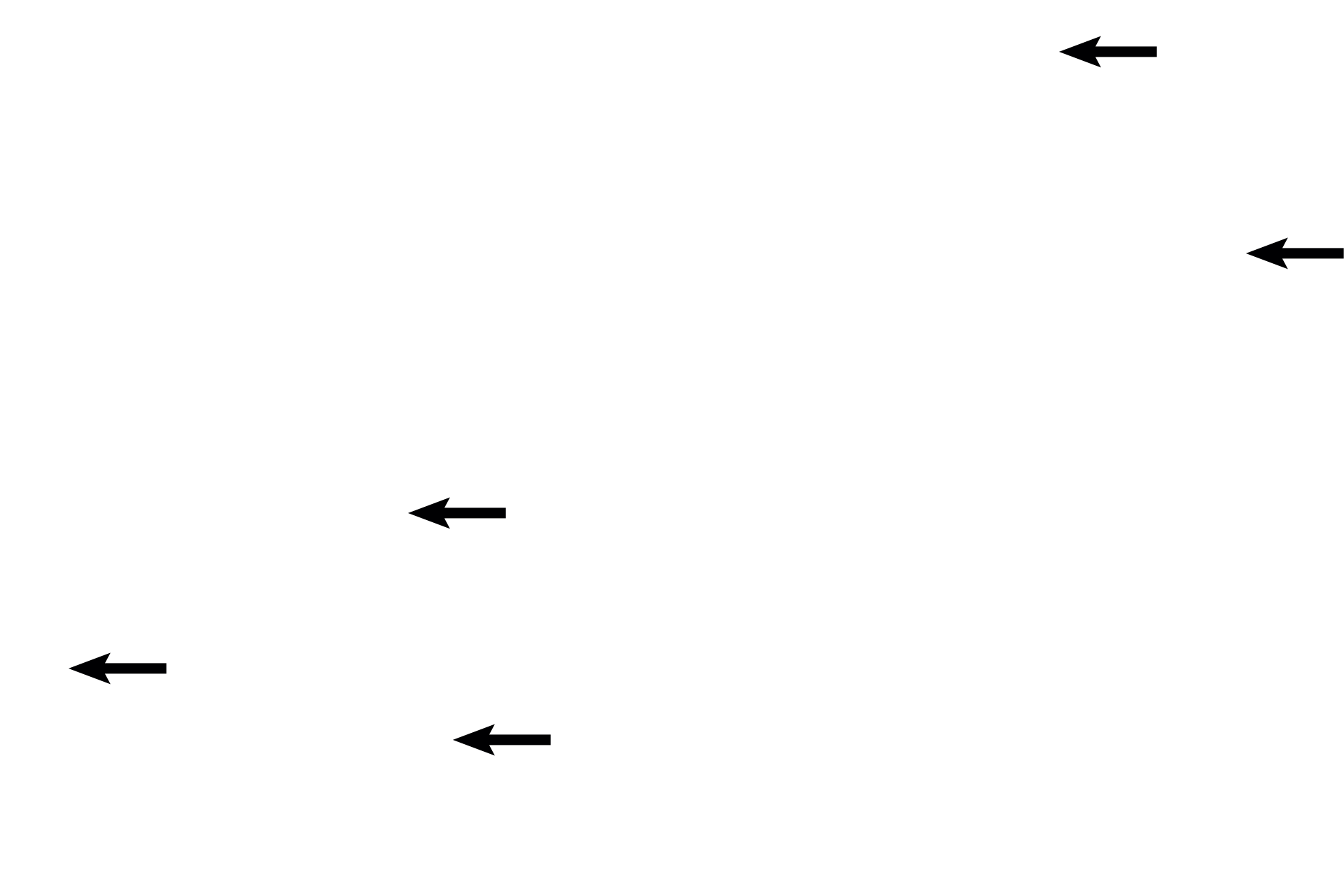
- Floating villi
Stem villi originate from the chorionic plate. Numerous, branching villi, termed floating villi, extend from each stem villus into the intervillous space, where they are bathed by maternal blood.
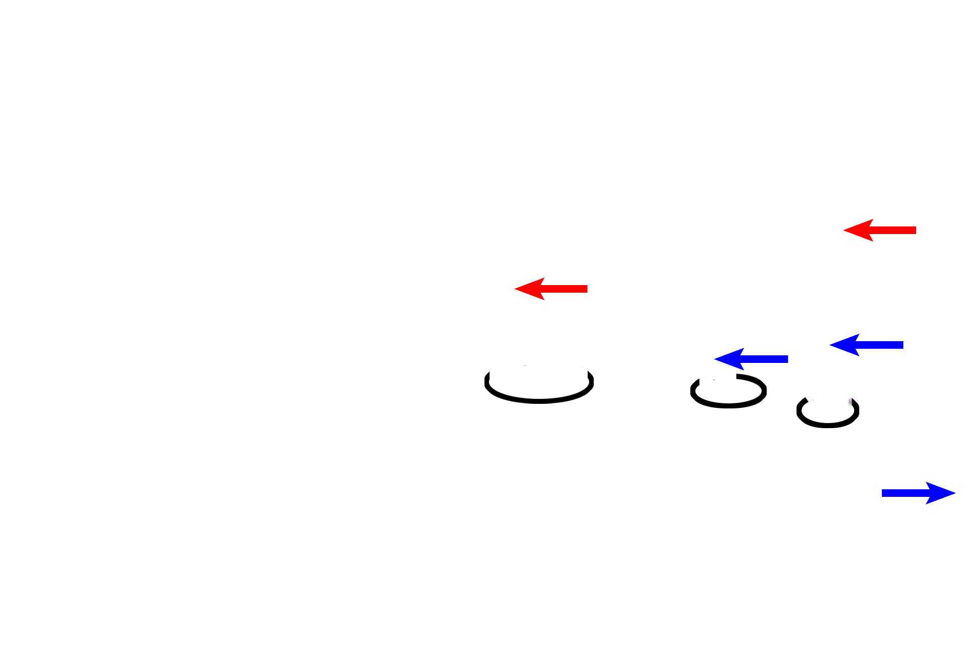
- Anchoring villi >
Anchoring villi (encircled), extend to the maternal side of the placenta, anchoring the thick, villous component to the trophoblastic shell and, indirectly, to the decidua. Embryonic CT (red arrows) does not extend all the way across the embryonic placenta; therefore, the central core of an anchoring villus is a column of cytotrophoblast cells (blue arrows) covered by syncytiotrophoblast.
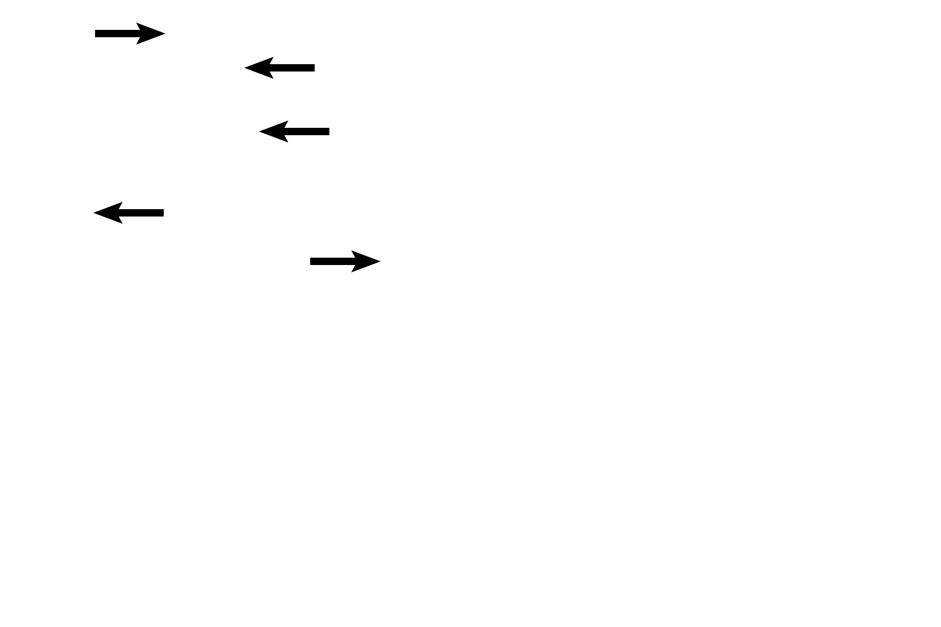
- Embryonic blood vessels >
Embryonic arteries pass through the chorionic plate and travel via stem villi into floating villi, where they form capillaries. Embryonic veins return this blood to the embryo. Maternal blood from the spiral arteries spurts into the intervillous space, bathing the floating villi with nutrient- and oxygen-rich blood. Embryonic blood vessels are difficult to differentiate in the right image.
Intervillous space
Embryonic arteries pass through the chorionic plate and travel via stem villi into floating villi, where they form capillaries. Embryonic veins return this blood to the embryo. Maternal blood from the spiral arteries spurts into the intervillous space, bathing the floating villi with nutrient- and oxygen-rich blood. Embryonic blood vessels are difficult to differentiate in the right image.
Trophoblastic shell >
The trophoblastic shell is a thick layer of cytotrophoblastic cells that forms the outer perimeter of the embryonic placenta. This layer still lies beneath the syncytiotrophoblast (blue arrows) and abuts the maternal decidua, sometimes separated from it by a layer of fibrinoid, an acidophilic extracellular material related to fibrin.

Fibrinoid
The trophoblastic shell is a thick layer of cytotrophoblastic cells that forms the outer perimeter of the embryonic placenta. This layer still lies beneath the syncytiotrophoblast (blue arrows) and abuts the maternal decidua, sometimes separated from it by a layer of fibrinoid, an acidophilic extracellular material related to fibrin.

Decidua >
The decidua, a layer of decidual cells derived from endometrial stromal cells, forms the maternal placenta. Present throughout the uterus, decidual regions are named according to their position relative to the embryo; the region seen here, lying beneath the functional embryonic placenta, is termed decidua basalis.

Next image >
The next image was taken from the embryonic side of the placenta in an area similar to that outlined by the rectangle.