
Glomerulus
This electron micrograph shows a podocyte, with its processes contacting three glomerular capillaries. A primary process of the podocyte branches to form secondary processes called pedicels, which rest on the glomerular basal lamina. Pedicels from one podocyte alternate with those from a different podocyte as they contact the capillary. Also visible is the fenestrated capillary endothelium. 6000x

Podocyte
This electron micrograph shows a podocyte, with its processes contacting three glomerular capillaries. A primary process of the podocyte branches to form secondary processes called pedicels, which rest on the glomerular basal lamina. Pedicels from one podocyte alternate with those from a different podocyte as they contact the capillary. Also visible is the fenestrated capillary endothelium. 6000x
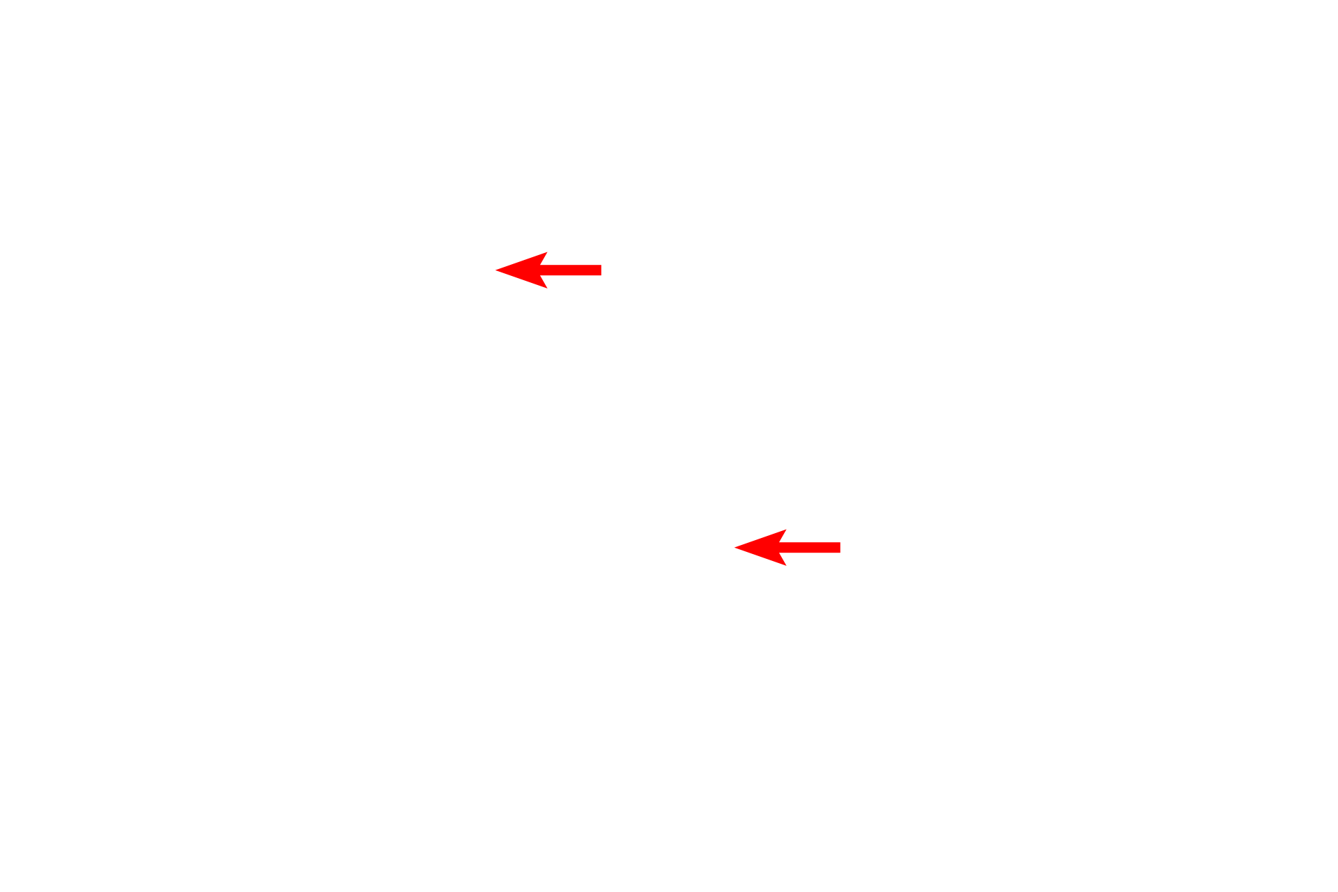
- Primary process
This electron micrograph shows a podocyte, with its processes contacting three glomerular capillaries. A primary process of the podocyte branches to form secondary processes called pedicels, which rest on the glomerular basal lamina. Pedicels from one podocyte alternate with those from a different podocyte as they contact the capillary. Also visible is the fenestrated capillary endothelium. 6000x
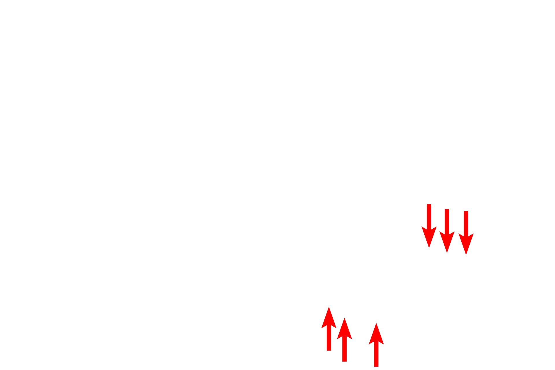
- Pedicels
This electron micrograph shows a podocyte, with its processes contacting three glomerular capillaries. A primary process of the podocyte branches to form secondary processes called pedicels, which rest on the glomerular basal lamina. Pedicels from one podocyte alternate with those from a different podocyte as they contact the capillary. Also visible is the fenestrated capillary endothelium. 6000x
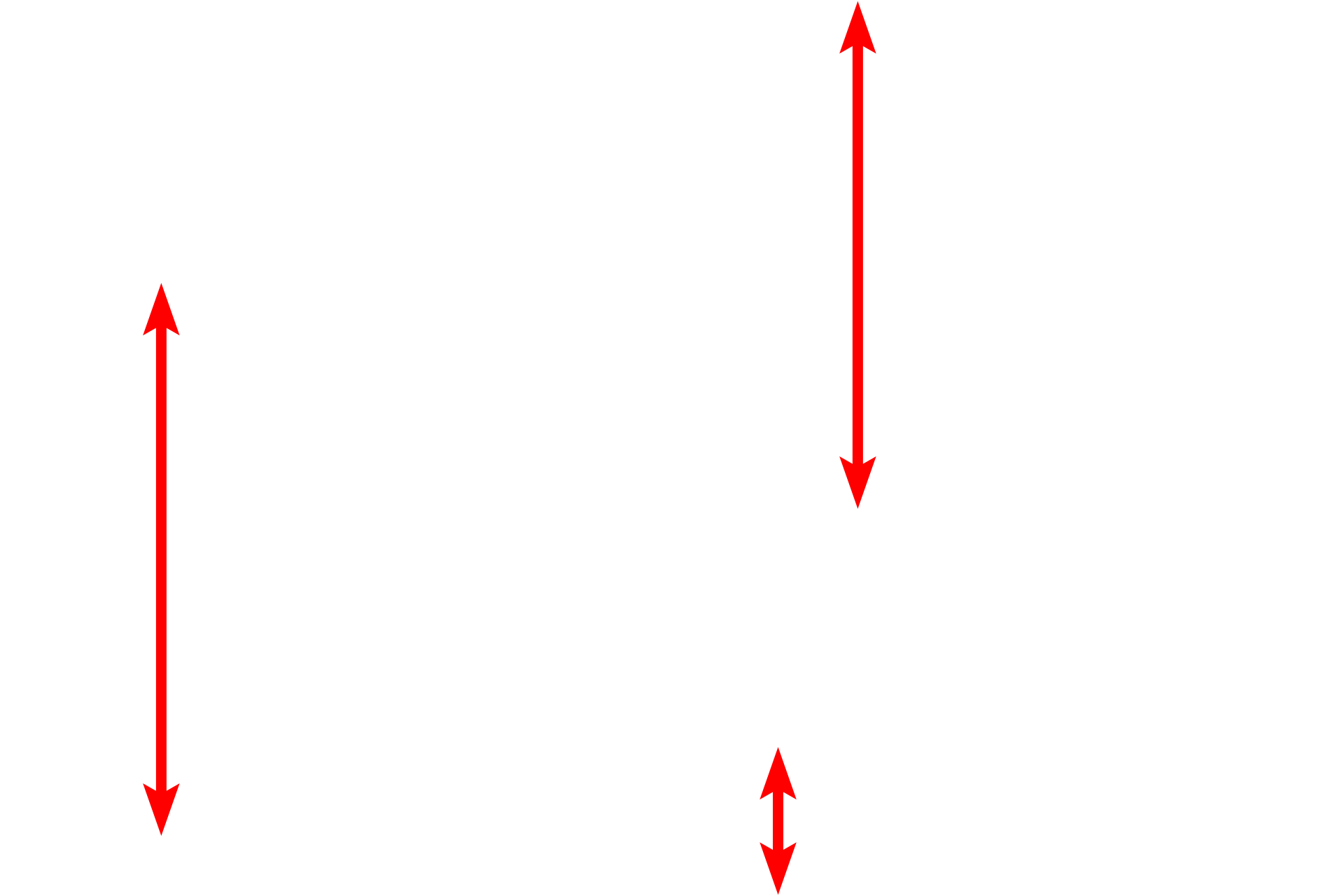
Glomerular capillaries
This electron micrograph shows a podocyte, with its processes contacting three glomerular capillaries. A primary process of the podocyte branches to form secondary processes called pedicels, which rest on the glomerular basal lamina. Pedicels from one podocyte alternate with those from a different podocyte as they contact the capillary. Also visible is the fenestrated capillary endothelium. 6000x
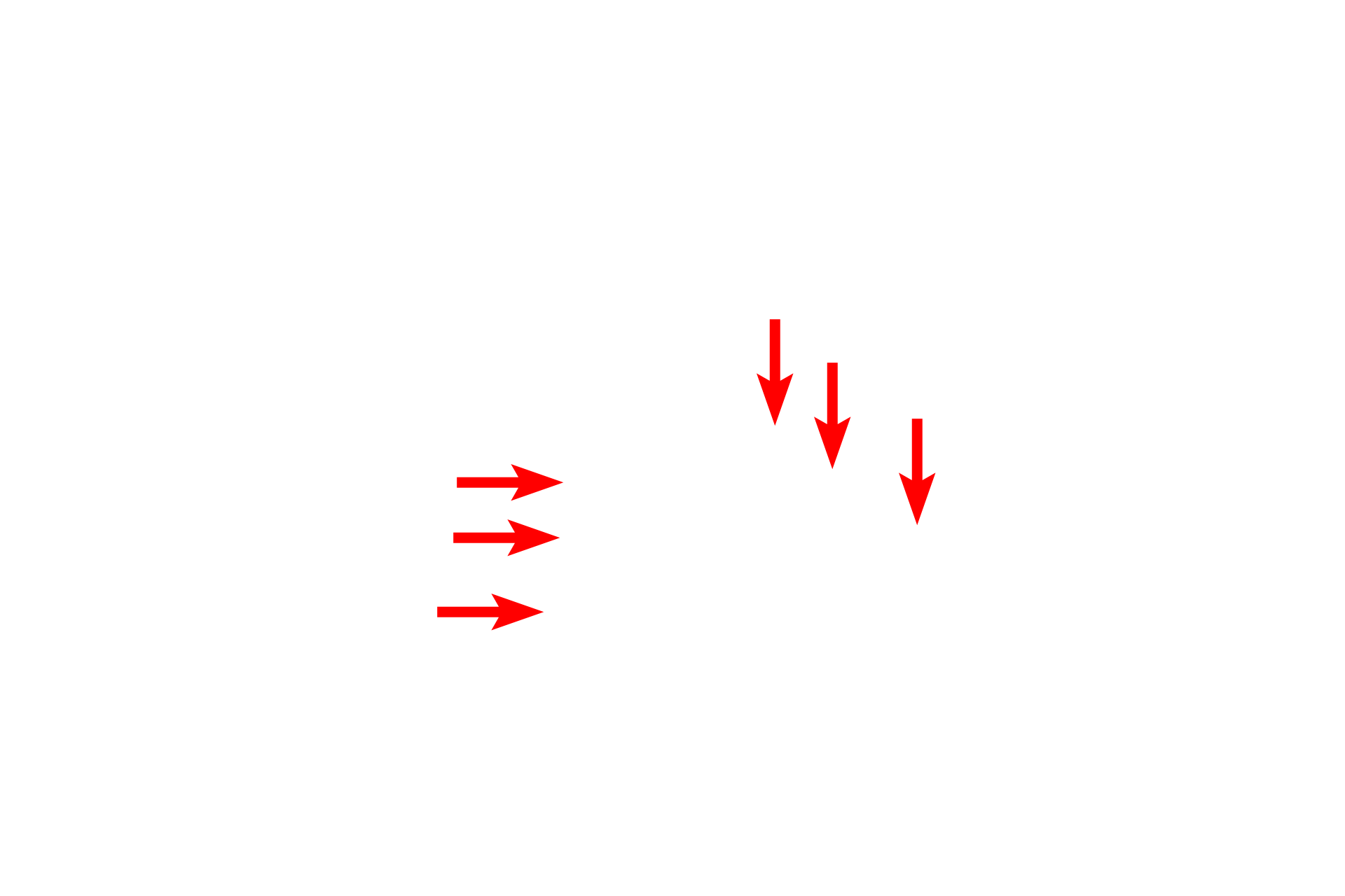
- Fenestrated endothelium
This electron micrograph shows a podocyte, with its processes contacting three glomerular capillaries. A primary process of the podocyte branches to form secondary processes called pedicels, which rest on the glomerular basal lamina. Pedicels from one podocyte alternate with those from a different podocyte as they contact the capillary. Also visible is the fenestrated capillary endothelium. 6000x
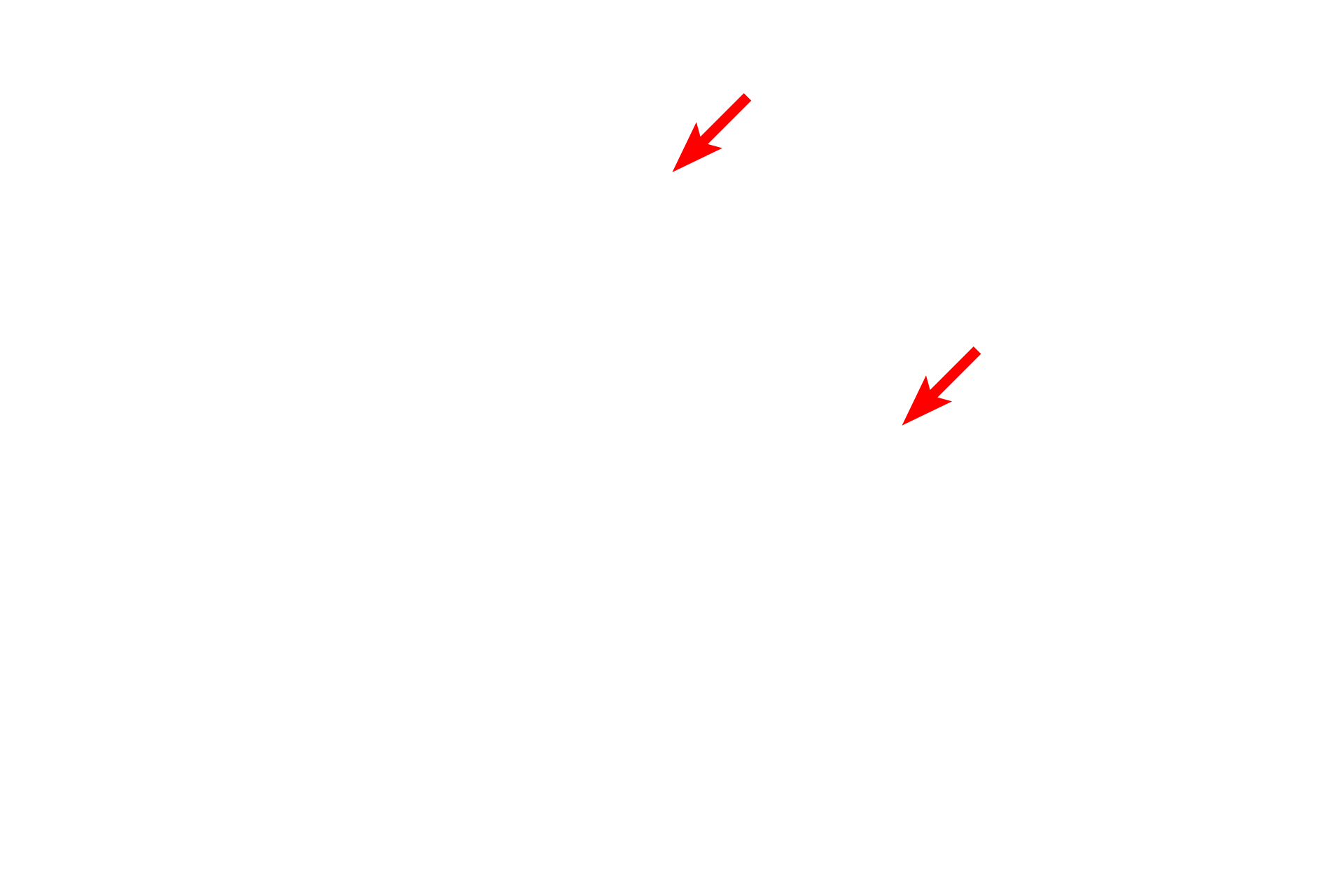
- Red blood cells
This electron micrograph shows a podocyte, with its processes contacting three glomerular capillaries. A primary process of the podocyte branches to form secondary processes called pedicels, which rest on the glomerular basal lamina. Pedicels from one podocyte alternate with those from a different podocyte as they contact the capillary. Also visible is the fenestrated capillary endothelium. 6000x
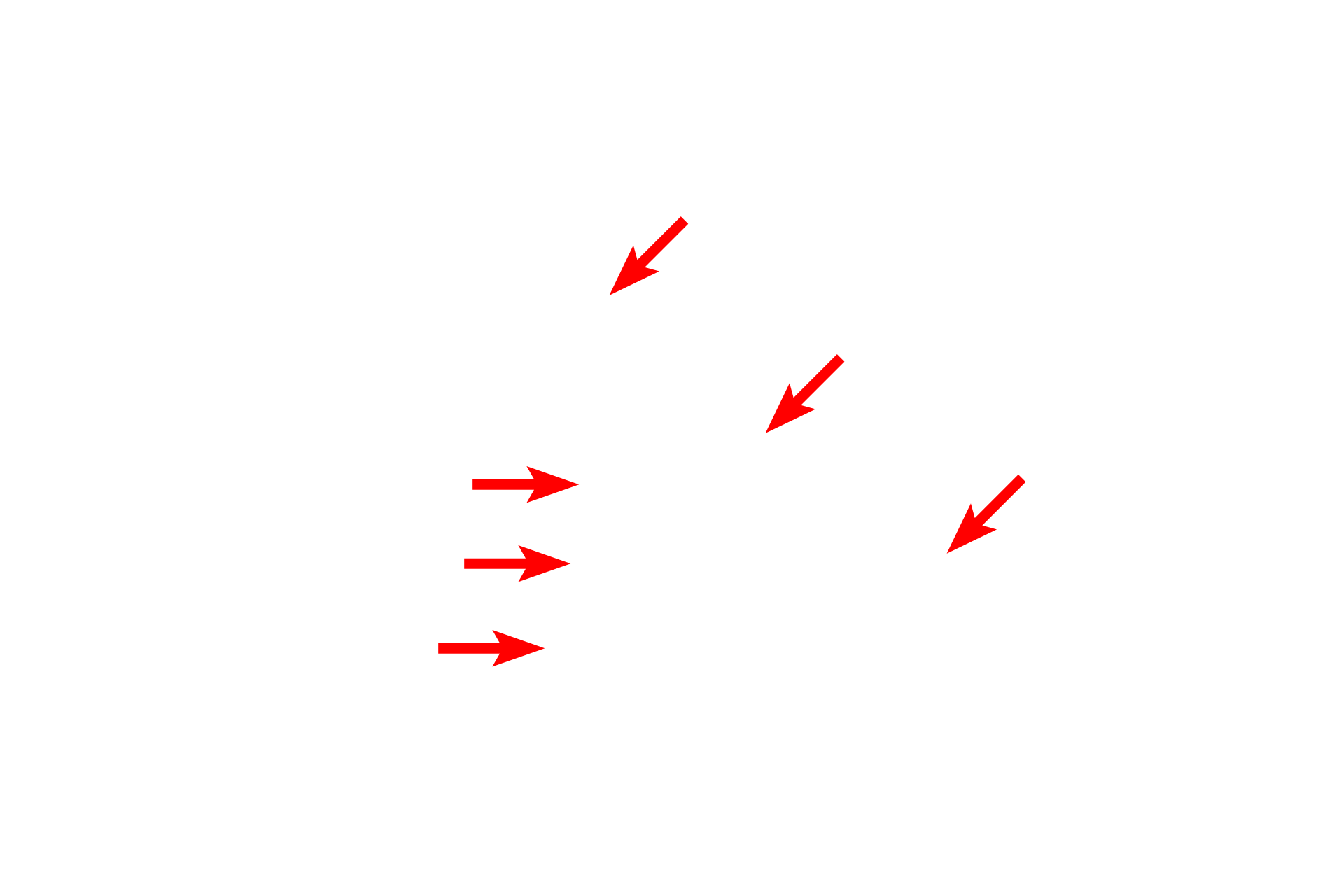
Basal laminae >
The thick basal lamina is formed by fusion of the individual podocyte and endothelial cell basal laminae. This layer plays a critical role in the glomerular filtration process as fluid leaves the capillary lumen and enters Bowman’s space.
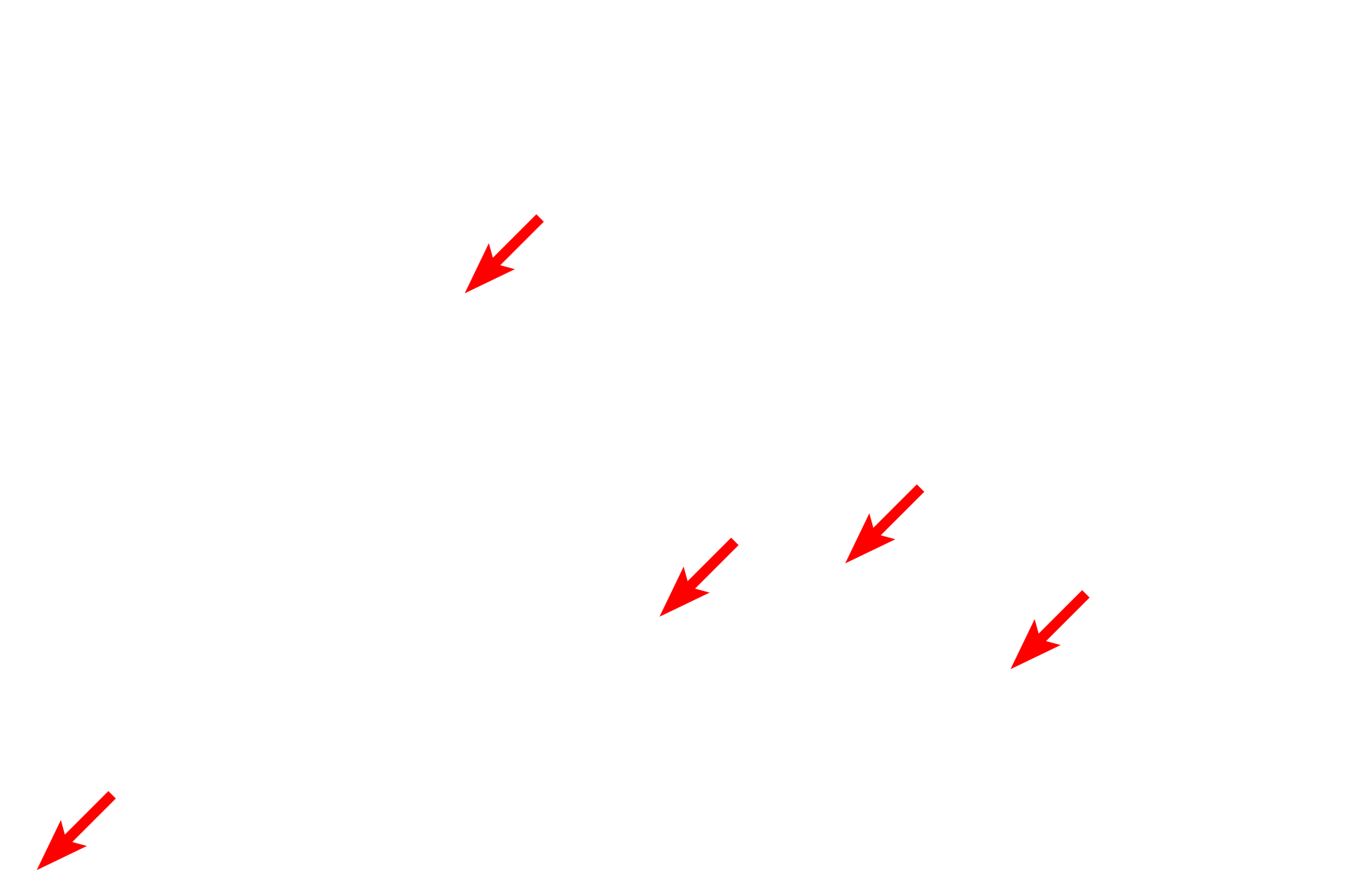
Bowman’s space
The thick basal lamina is formed by fusion of the individual podocyte and endothelial cell basal laminae. This layer plays a critical role in the glomerular filtration process as fluid leaves the capillary lumen and enters Bowman’s space.
 PREVIOUS