
Endochondral ossification
The epiphyseal plate, located between the primary and secondary centers of ossification, is comprised of zones of cartilage, reflecting stages of chondrocyte regression. Calcified cartilage spicules formed by this process serve as a framework on which bone is deposited. Such bone replaces previously existing cartilage and, thus, is formed endochondrally. 200x
Resting zone >
The resting zone is composed of typical hyaline cartilage that serves as a reserve cell source for the epiphyseal plate.
Zone of proliferation >
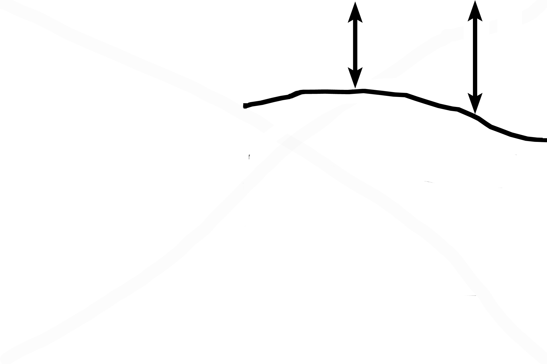
Chondrocytes in the zone of proliferation undergo interstitial growth to maintain the thickness of the plate and well as advancing the epiphysis upwards. Instead of forming spherical isogenous groups, these dividing cells become aligned in rows (arrows), a pattern that results in vertical columns of cartilage matrix between them. Such an orientation guarantees that the calcified cartilage spicules remaining after chondrocyte death will be linearly arranged.

Zones of maturation-hypertrophy-calcification >
Chondrocytes begin regressive changes during which cells mature, hypertrophy (increase in size) and begin secreting alkaline phosphatase into the matrix. This secretion results in calcification of the matrix. These zones are grouped here into a single zone, the zone of maturation, hypertrophy and calcification (Zones of M-H-C).

Zone of degeneration >
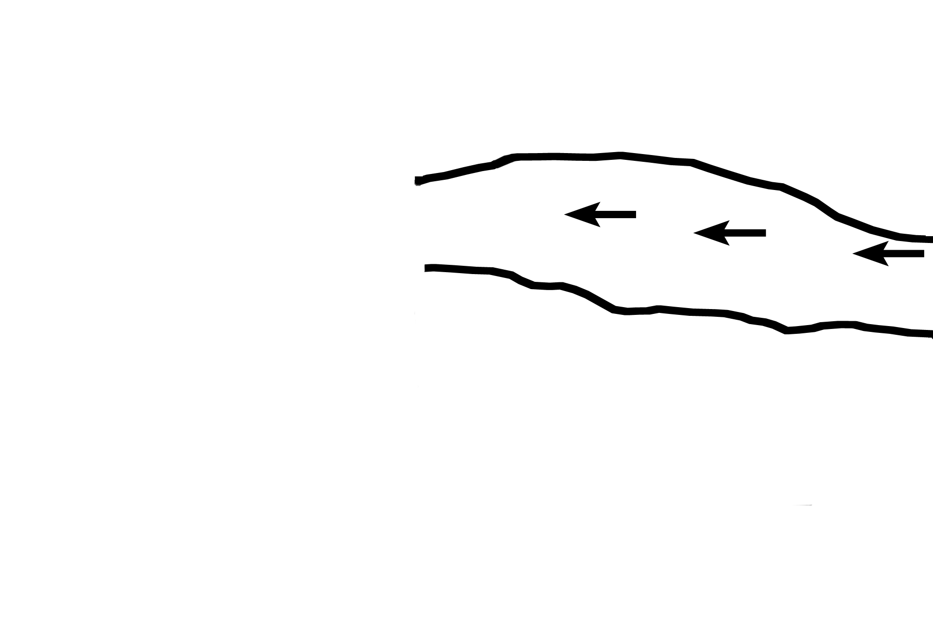
The zone of degeneration is formed as the calcified cartilage matrix prevents nutrients and oxygen from diffusing to the chondrocytes and their resultant degeneration. The calcified cartilage remaining between the columns of empty chondrocyte lacunae forms spicules on which bone will be deposited.
Zone of ossification >
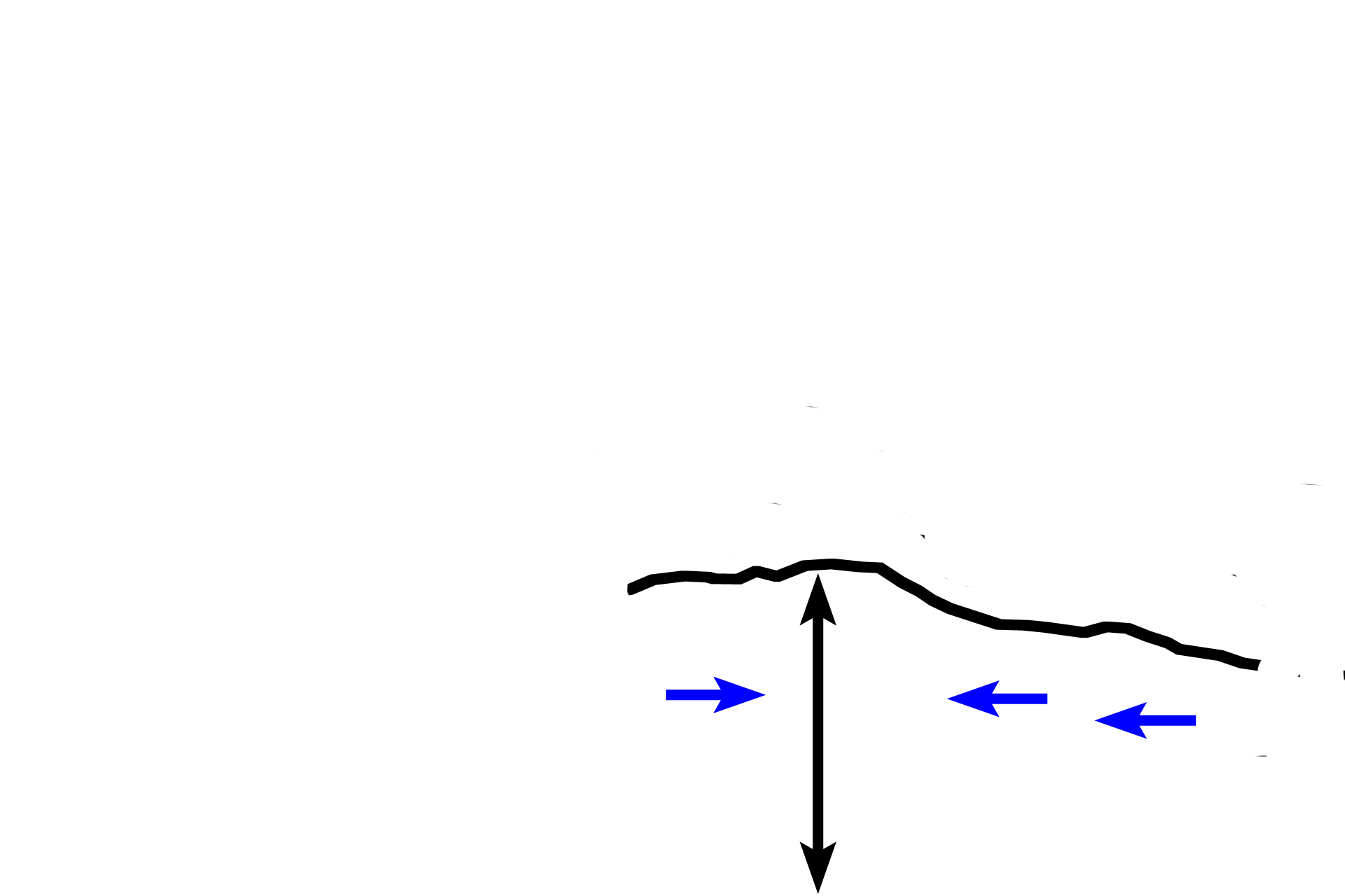
The zone of ossification is formed as osteoblasts deposit bone (blue arrows) on the calcified cartilage spicules. The addition of new bone to the ends of the periosteal collar, adds to the length of the diaphysis and elongation of the bone. The bone deposited in the center of the zone is resorbed to maintain the marrow cavity.

Zone of resorption >
This zone is not present in this section; it is located below the zone of ossification. The zone of resorption is formed by the breakdown of much of the newly formed bone/calcified cartilage spicules by osteoclasts. This resorption maintains the marrow cavity.

Periosteal collar >
The periosteal collar (arrows) forms the diaphysis of the long bone. The band is a cylinder of bone laid down intramembranously on the surface of the hyaline cartilage template by the periosteum. Growth of a bone in length occurs by addition of bone to the ends of the periosteal collar by endochondral ossification.

Periosteum
The periosteal collar (arrows) forms the diaphysis of the long bone. The band is cylinder of bone laid down intramembranously on the surface of the hyaline cartilage template by the periosteum. Growth of a bone in length occurs by addition of bone to the ends of the periosteal collar by endochondral ossification.

Next images
The next three images were taken from areas similar to those outlined by the rectangles.