Peripheral nerve
The endoneurium surrounds Schwann cells and the axons they ensheath. This peripheral nerve contains both myelinated and unmyelinated axons, which are surrounded by a perineurium to form a fascicle. This tissue was fixed with osmium to preserve lipid and, thus, the myelin sheaths are well preserved. 1500x

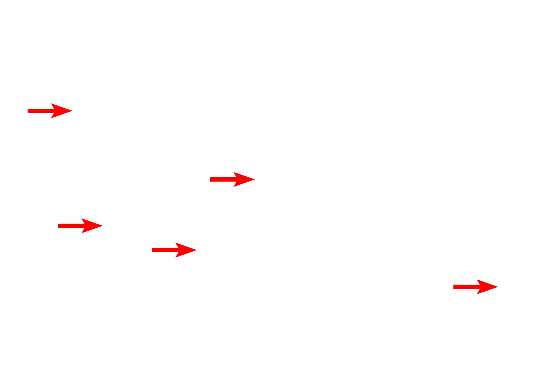
Myelinated axons >
Axons in the peripheral nervous system that possess a myelin sheath are usually larger than one micron in diameter. The thickness of the myelin sheath is proportional to the diameter of the axon. Myelin is composed of concentric wrappings of the Schwann cell plasma membrane. Each Schwann cell associates with one axon to produce a single internodal segment of myelin.

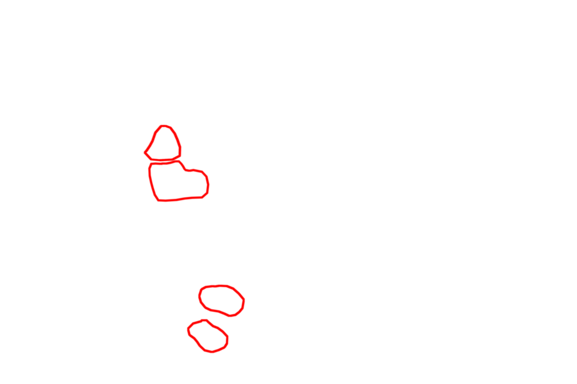
Unmyelinated axons >
Unmyelinated axons are generally less than one micron in diameter. Unlike myelinated axons, multiple axons are associated with a single Schwann cell (circled in red); each axon is embedded in a longitudinal indentation of the Schwann cell surface. Each of the numerous unmyelinated axons, resembling a white dot, is just resolvable at this magnification within the outlines.

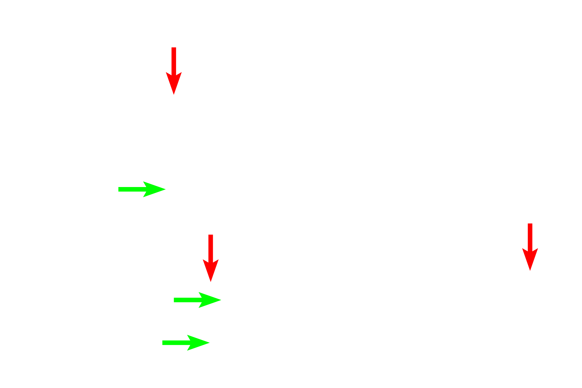
Schwann cell nuclei >
The Schwann cell nuclei are seen in association with individual myelinated axons (red arrows), as well as clusters of unmyelinated axons (green arrows). Each myelin-forming Schwann cell forms a single internode of myelin around one axon, whereas one non-myelinating Schwann cell envelopes multiple, small-caliber axons. Each Schwann cell also secretes its own external lamina.

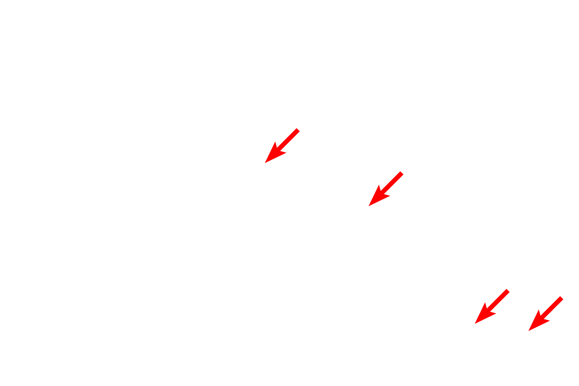
Endoneurium >
The endoneurium is composed of fibroblasts and collagen fibrils interspersed between the Schwann cells and the axons they ensheath.

Perineurium >
The perinerium consists of one or more layers of squamous cells which form the protective blood-nerve barrier. These highly unique cells exhibit epithelial features, e.g., basal laminae and tight junctions; however, they are also contractile, possessing large numbers of actin filaments, and synthesize collagen, thus resembling smooth muscle cells and fibroblasts.

Fibroblast
The Schwann cell nuclei are seen in association with individual myelinated axons (red arrows), as well as clusters of unmyelinated axons (green arrows). Each myelin-forming Schwann cell forms a single internode of myelin around one axon, whereas one non-myelinating Schwann cell envelopes multiple, small-caliber axons. Each Schwann cell also secretes its own external lamina.
 PREVIOUS