
Skeletal muscle
This H&E section shows muscle fibers cut in cross section and clearly demonstrates the myofibrils that fill the muscle fibers. Numerous capillaries are also visible. This is a section of the tongue which contains interlacing skeletal muscle fascicles and thus at the top and bottom of the image are fibers cut longitudinally. 800x
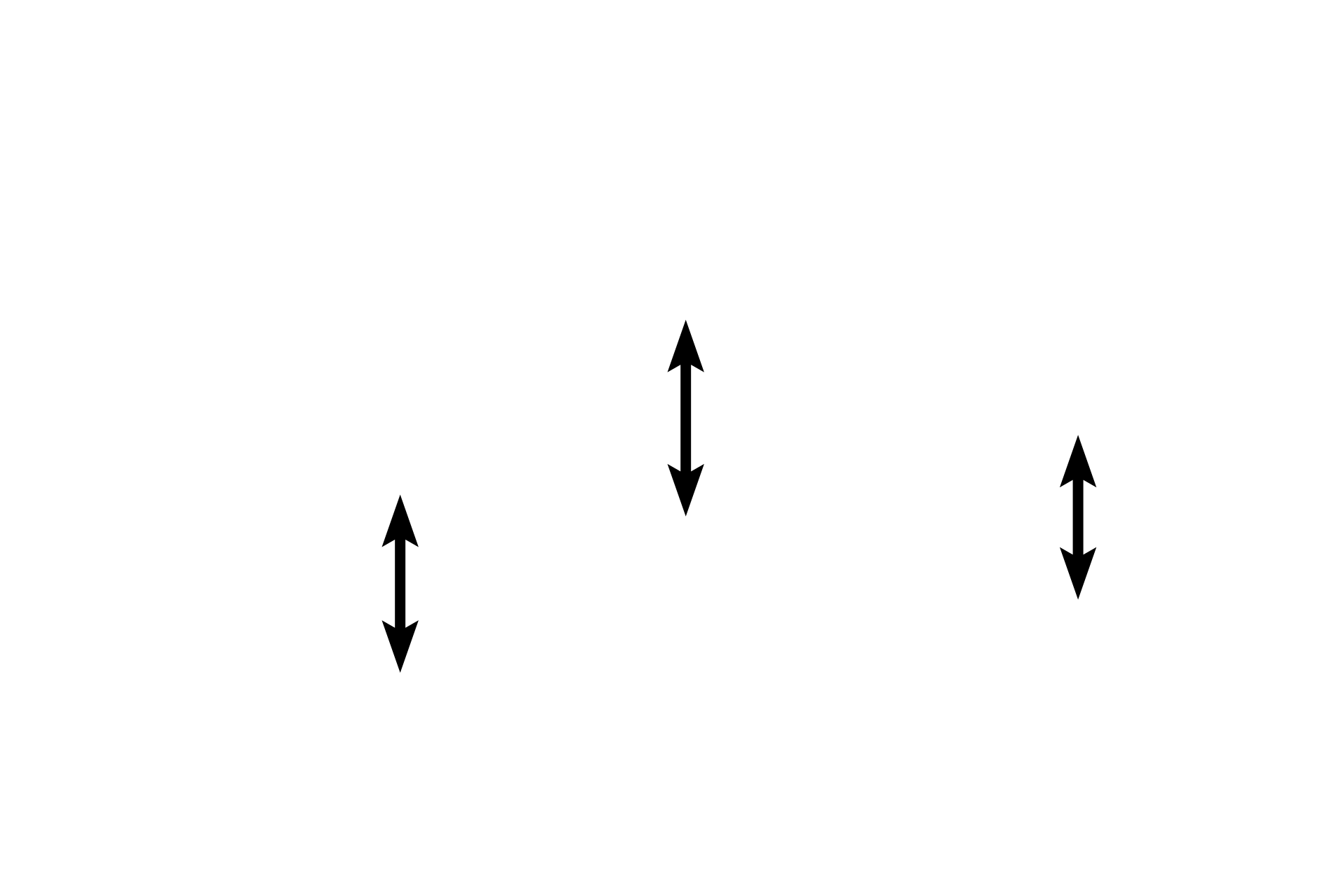
Muscle fibers
This H&E section shows muscle fibers cut in cross section and clearly demonstrates the myofibrils that fill the muscle fibers. Numerous capillaries are also visible. This is a section of the tongue which contains interlacing skeletal muscle fascicles and thus at the top and bottom of the image are fibers cut longitudinally. 800x
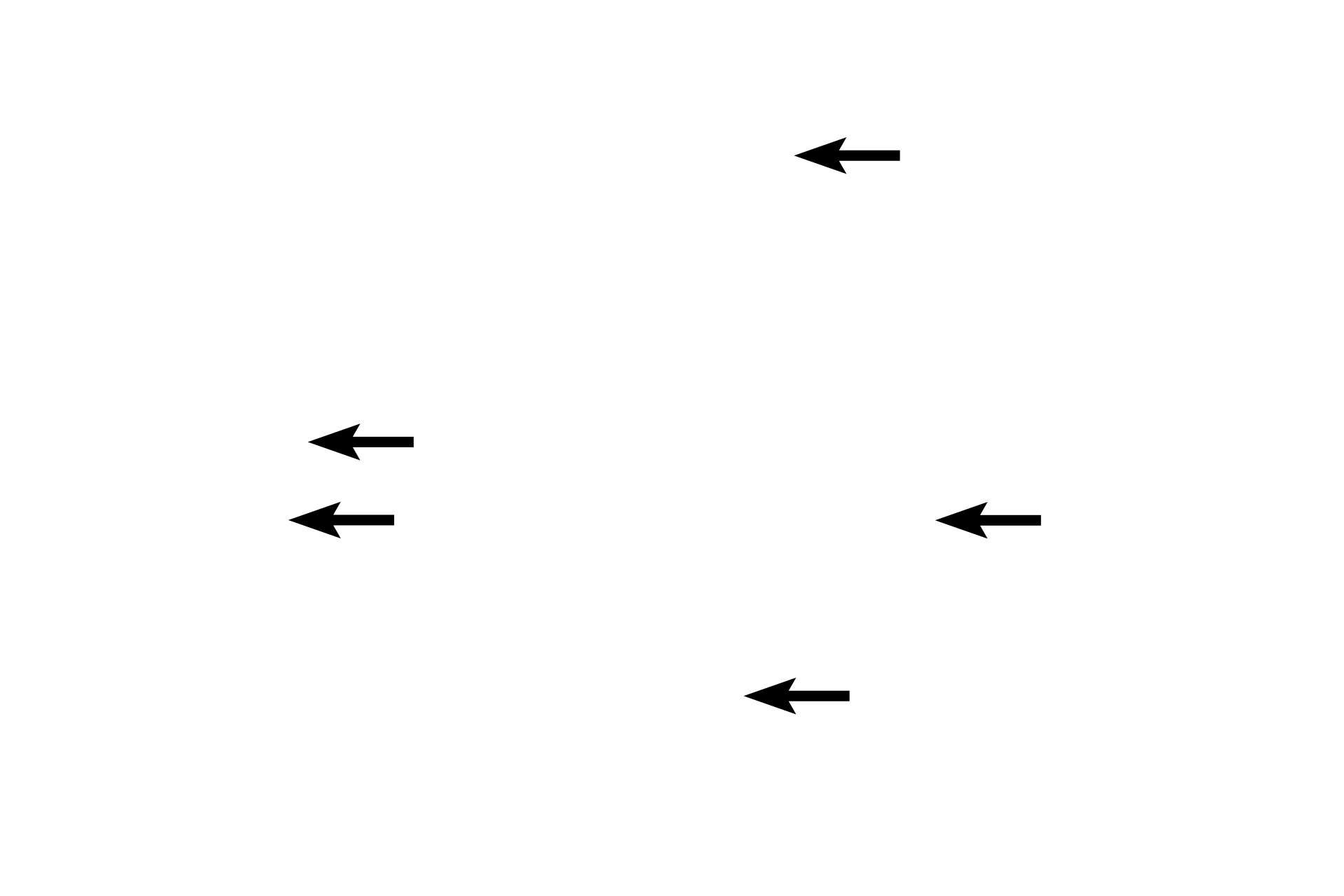
Myofibrils
This H&E section shows muscle fibers cut in cross section and clearly demonstrates the myofibrils that fill the muscle fibers. Numerous capillaries are also visible. This is a section of the tongue which contains interlacing skeletal muscle fascicles and thus at the top and bottom of the image are fibers cut longitudinally. 800x
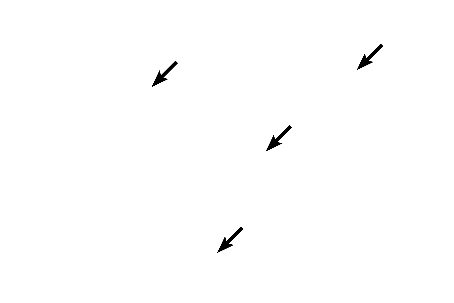
Nuclei
This H&E section shows muscle fibers cut in cross section and clearly demonstrates the myofibrils that fill the muscle fibers. Numerous capillaries are also visible. This is a section of the tongue which contains interlacing skeletal muscle fascicles and thus at the top and bottom of the image are fibers cut longitudinally. 800x
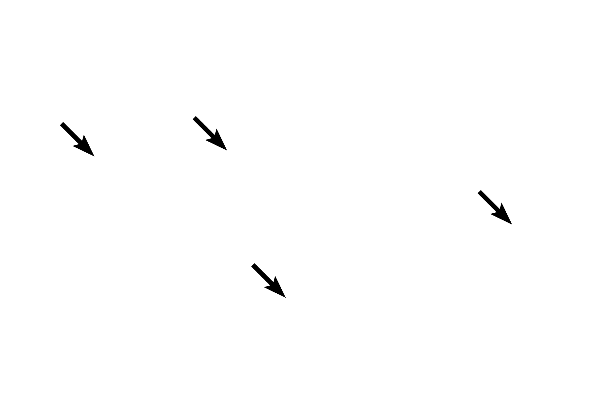
Capillaries
This H&E section shows muscle fibers cut in cross section and clearly demonstrates the myofibrils that fill the muscle fibers. Numerous capillaries are also visible. This is a section of the tongue which contains interlacing skeletal muscle fascicles and thus at the top and bottom of the image are fibers cut longitudinally. 800x
Endomysium
This H&E section shows muscle fibers cut in cross section and clearly demonstrates the myofibrils that fill the muscle fibers. Numerous capillaries are also visible. This is a section of the tongue which contains interlacing skeletal muscle fascicles and thus at the top and bottom of the image are fibers cut longitudinally. 800x
Image source >
This image is taken of a slide in the University of Mississippi slide collection.
 PREVIOUS