
Skeletal muscle
At high magnification, myofibrils in muscle fibers are clearly visible. Myofibrils are composed of myofilaments, consisting mostly of the proteins actin and myosin. Myofilaments cannot be resolved individually with the light microscope. The dark, wavy line around each fiber represents both the sarcolemma and endomysium. 1000x
Muscle fibers >
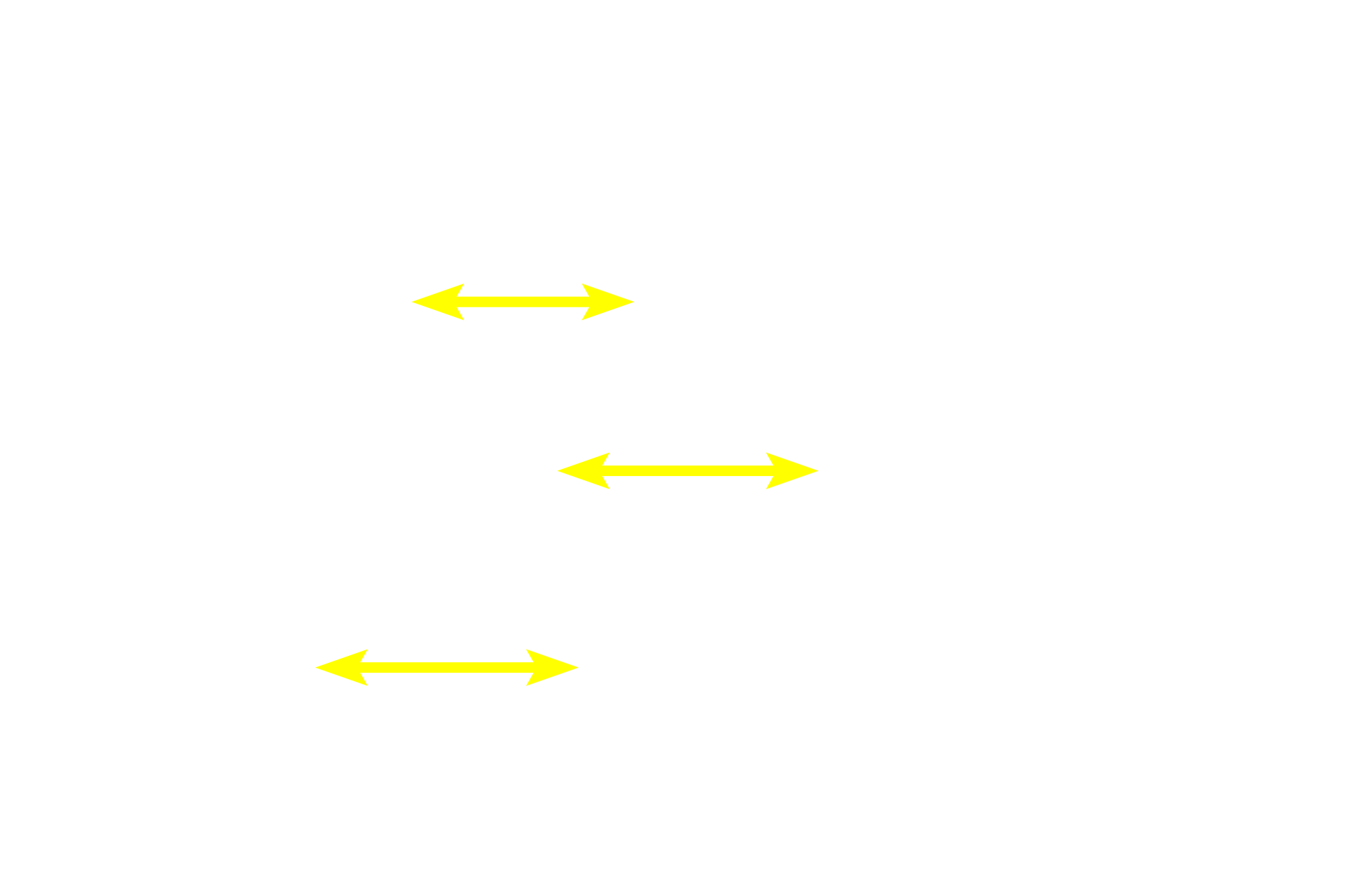
Skeletal muscle fibers constitute the largest muscle fiber type. They are long, uniform cylinders with multiple, peripheral nuclei. Developmentally, these fibers form by the fusion of many myoblasts, producing a very large, single cell with multiple nuclei. Such a cell is referred to as a syncytium. Myofibrils containing the contractile myofilaments fill the cytoplasm.
- Myofibrils >
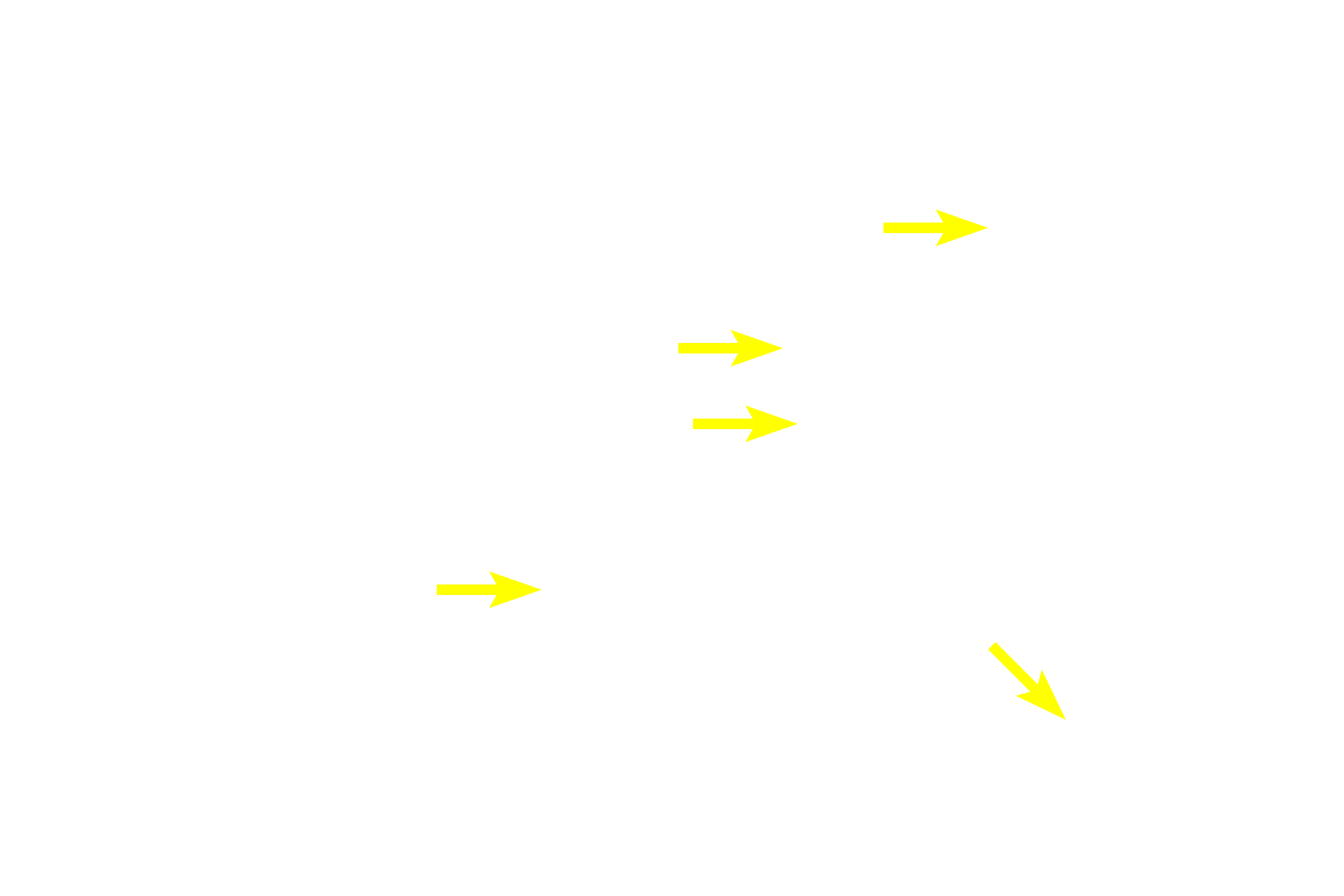
Myofibrils run parallel to the longitudinal axis of the fiber and, therefore, are cut in cross section here. Myofibrils occupy most of the cytoplasm of the cell while other organelles, like mitochondria and ER, are located between them. Multiple nuclei are located peripherally, directly beneath the sarcolemma.
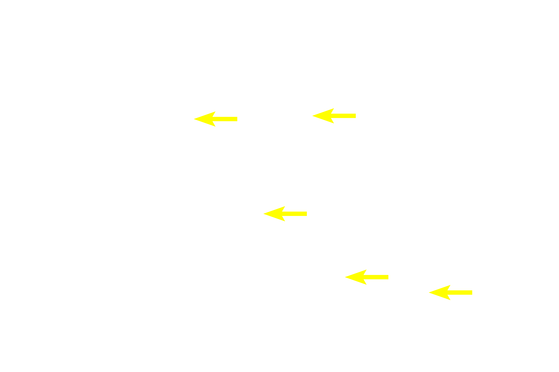
- Nuclei >
Multiple nuclei are located at the periphery of each muscle fiber.
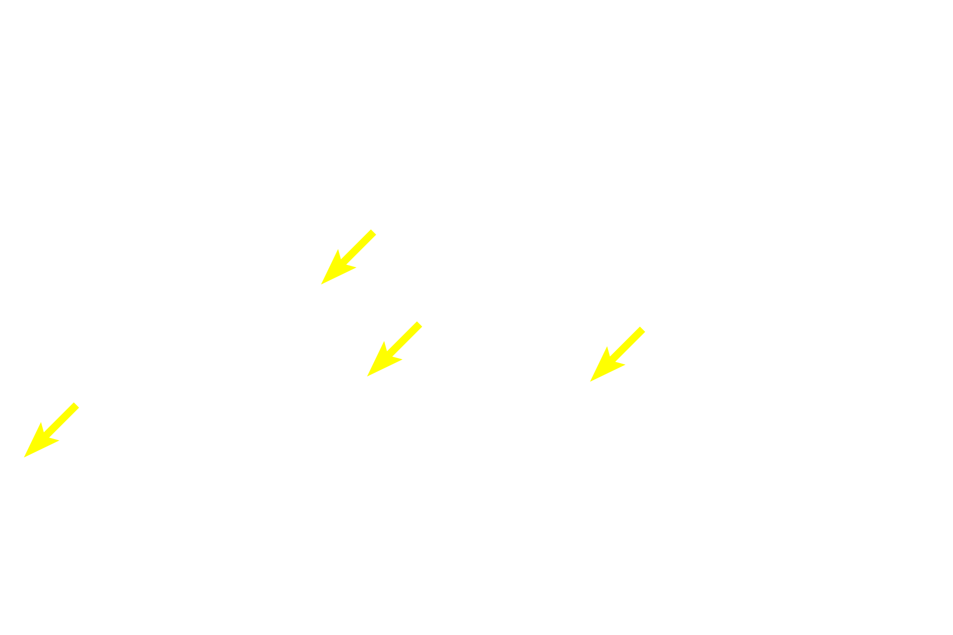
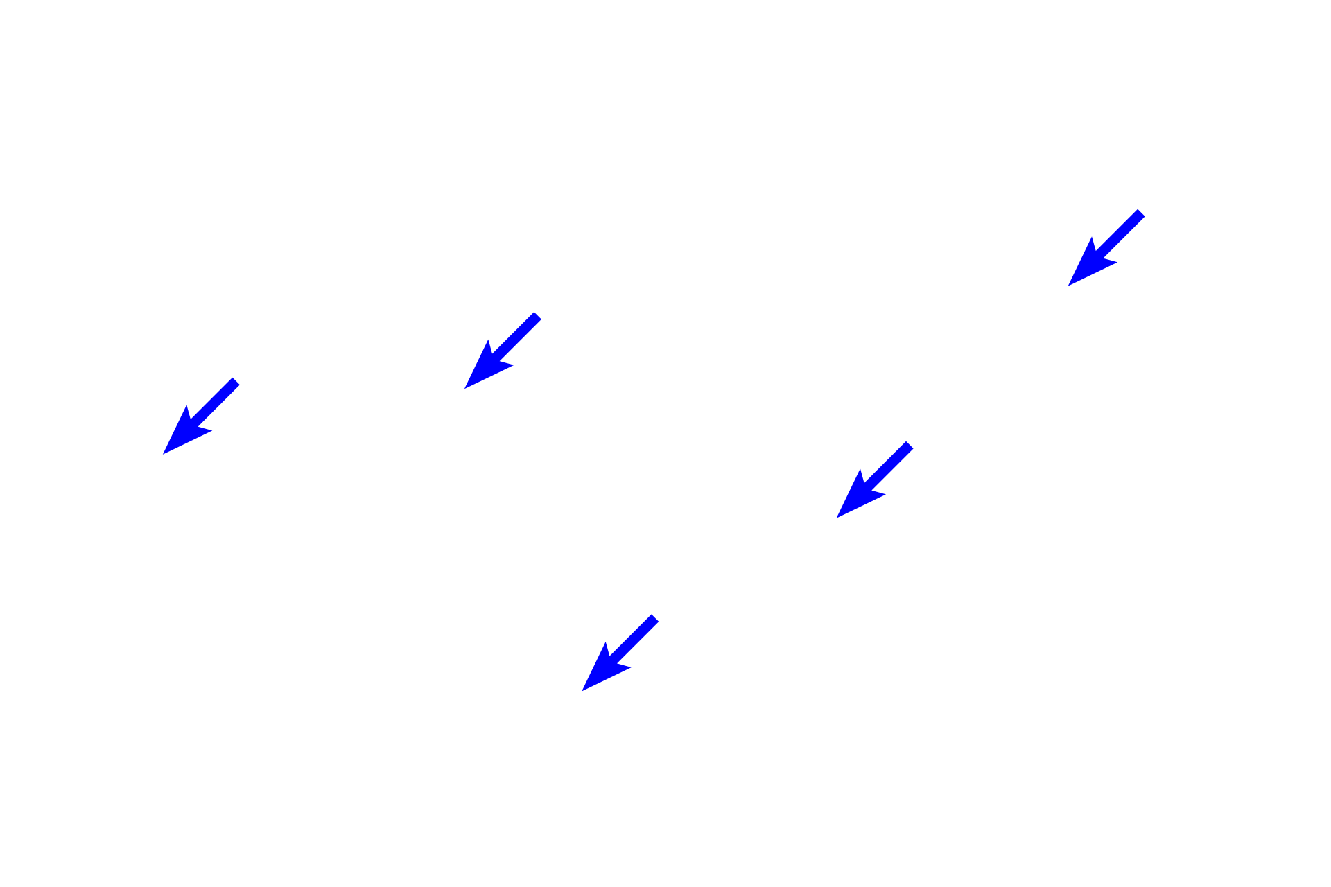
Blood vessels >
Due to its high metabolic demand, skeletal muscle is well vascularized.
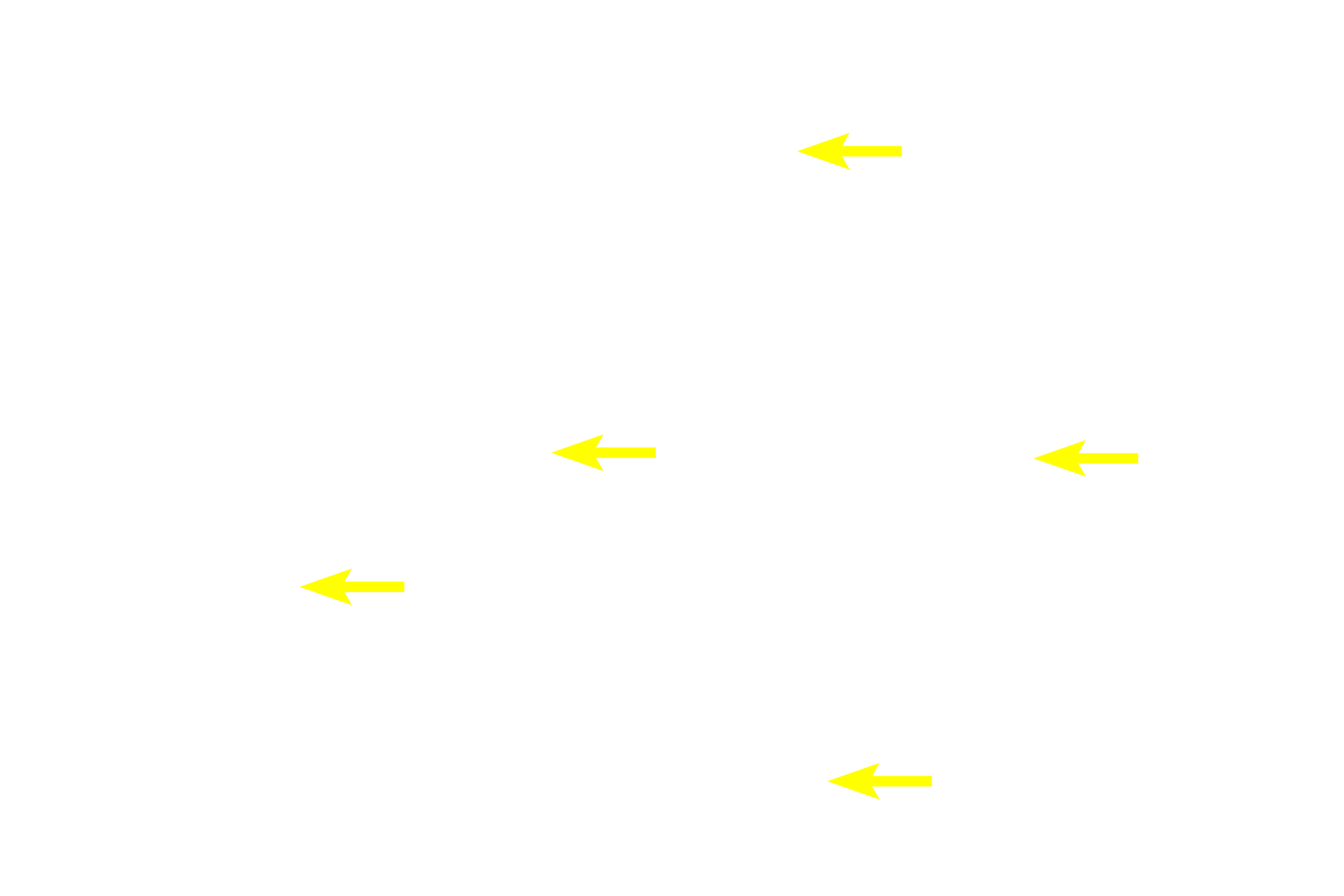
Endomysium >
The endomysium consists of a meshwork of reticular fibers that surround individual muscle fibers like a sleeve.
Artifactual space
Clear spaces in the image are the result of shrinkage during tissue processing.