
Skeletal muscle
A Masson-stained cross section of skeletal muscle shows clearly the association of connective tissue with the muscle fibers. An endomysium of reticular fibers surrounds individual muscle fibers, which are filled with myofibrils. A portion of the perimysium is visible in the lower right. 400x
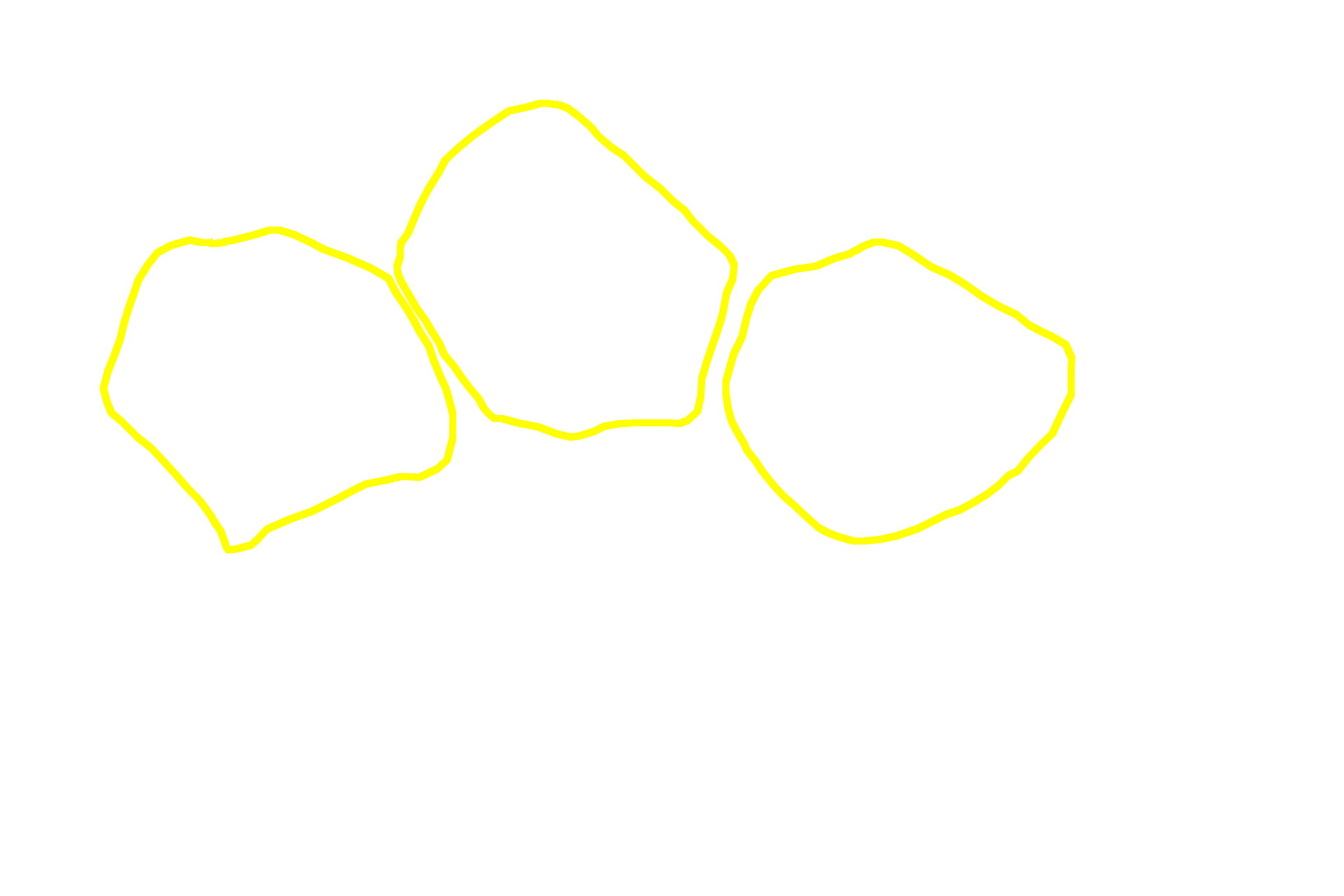
Muscle fibers >
Muscle fibers appear as irregular polygons and their cytoplasm is filled with myofibrils. Their nuclei are not visible in this image.
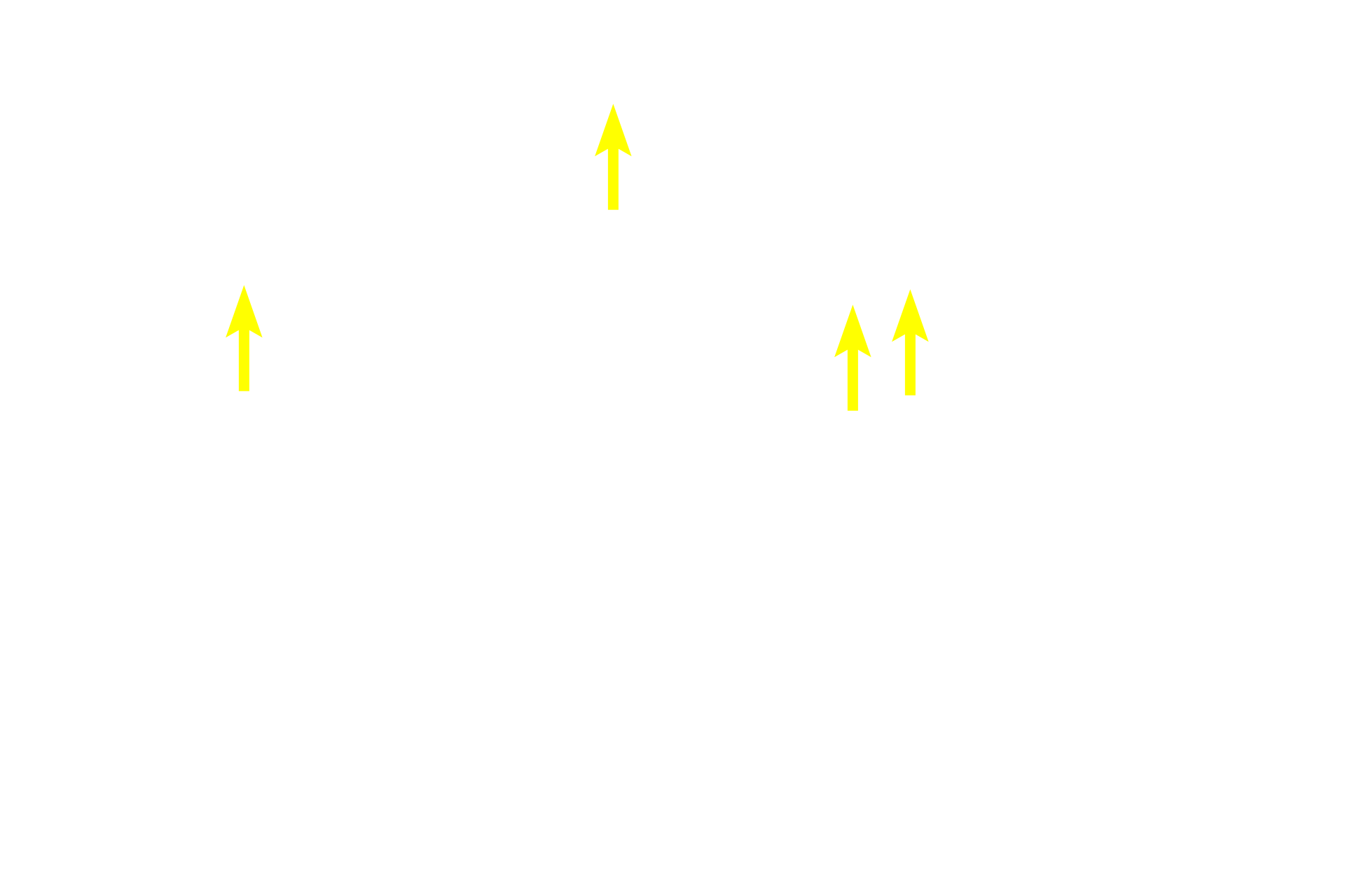
- Myofibrils
Muscle fibers appear as irregular polygons and their cytoplasm is filled with myofibrils. Their nuclei are not visible in this image.
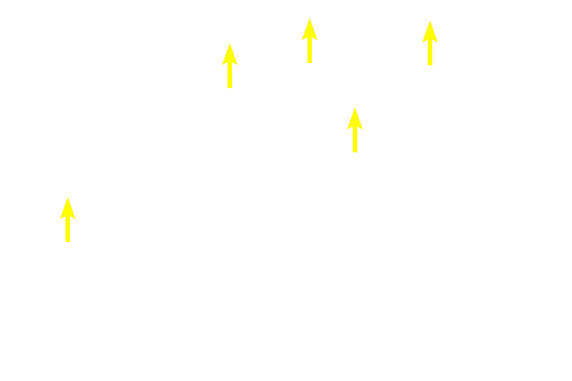
Endomysium >
The endomysium consists of a meshwork of reticular fibers that surround individual muscle fibers like a sleeve. In addition, the muscle fibers themselves secrete an external lamina that is similar in composition to the basal lamina of epithelial tissues. The external lamina is closely apposed to the endomysium and some consider it to be a component of the endomysium.
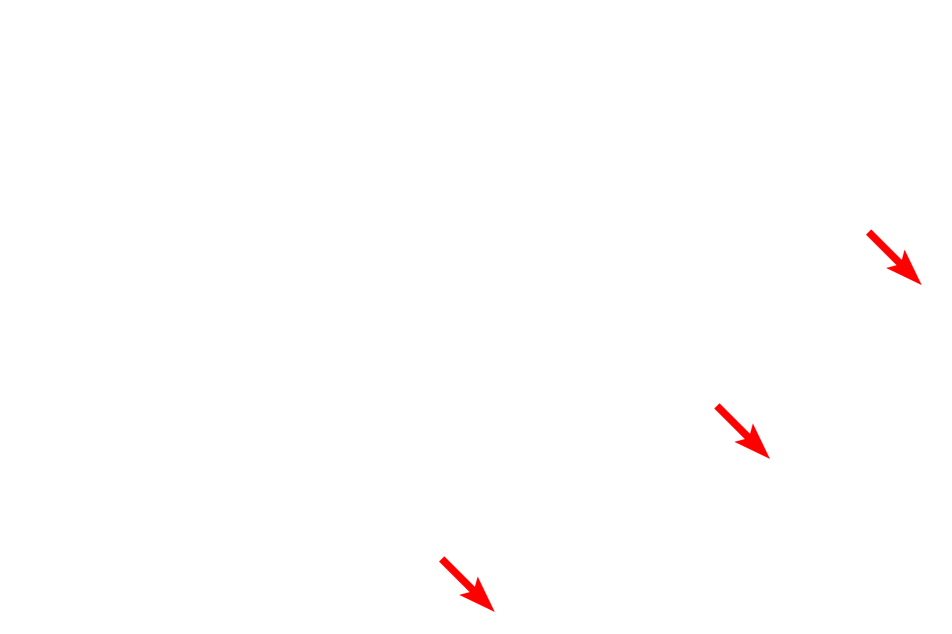
Perimysium >
The perimysium, composed of Type I collagen fibers, encloses groups of skeletal muscle fibers, forming functional groups called fascicles. Larger blood vessels and nerves travel in the perimysium.

Next image >
The electron micrograph in the next image shows the region in the rectangle.
 PREVIOUS