
Cardiac muscle
A high magnification electron micrograph shows portions of four myofibrils. This banding pattern and the changes that occur during contraction are the same as those for skeletal muscle fibers. Note the precise alignment of the sarcomeres. 40,000x
Myofibrils
A high magnification electron micrograph shows portions of four myofibrils. This banding pattern and the changes that occur during contraction are the same as those for skeletal muscle fibers. Note the precise alignment of the sarcomeres. 40,000x
Sarcomeres
A high magnification electron micrograph shows portions of four myofibrils. This banding pattern and the changes that occur during contraction are the same as those for skeletal muscle fibers. Note the precise alignment of the sarcomeres. 40,000x
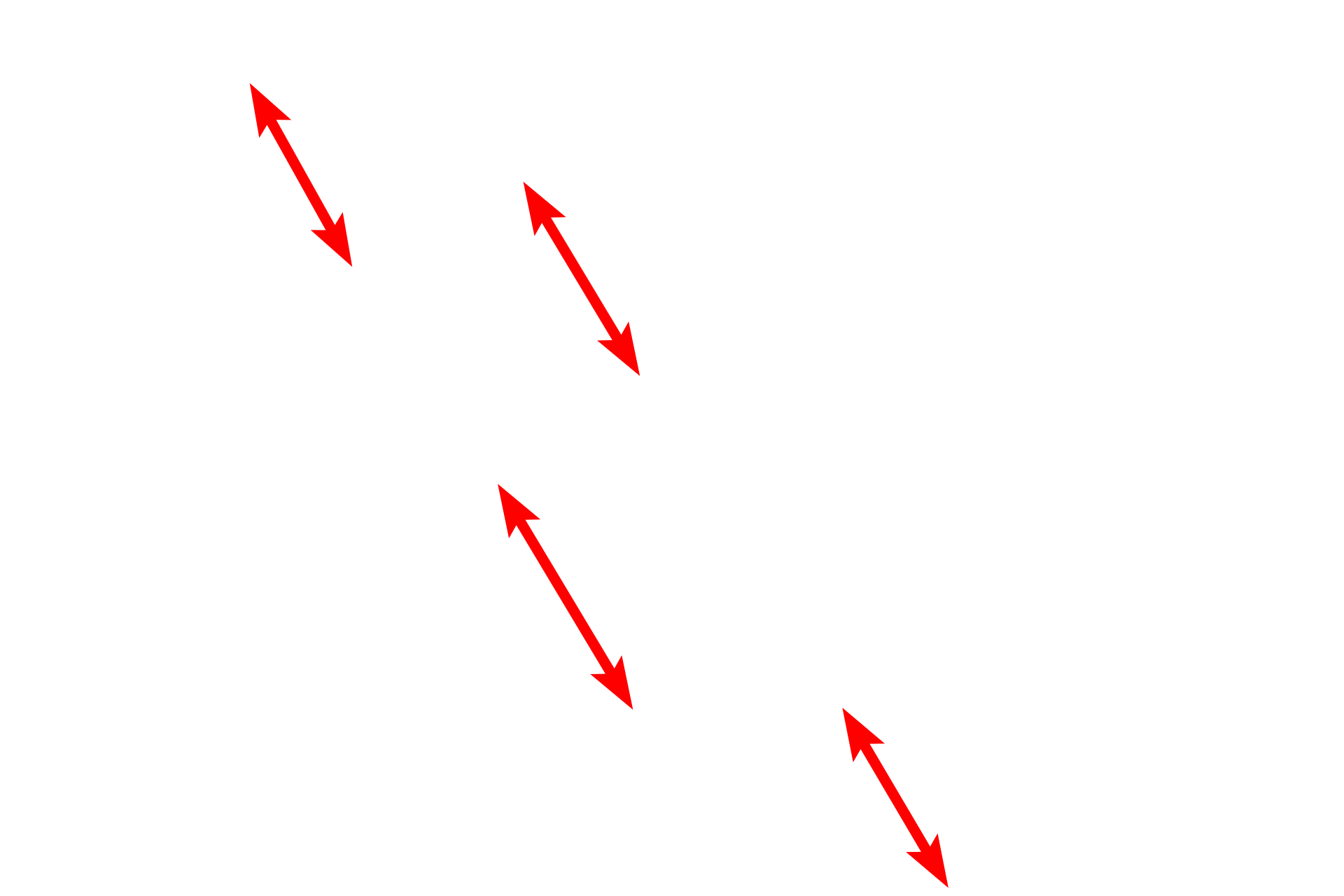
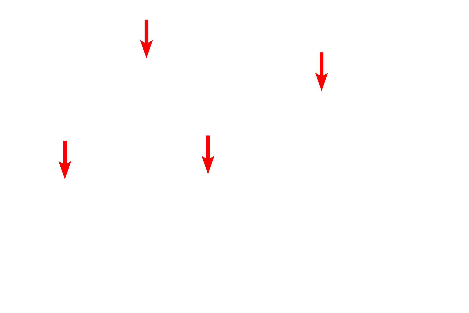
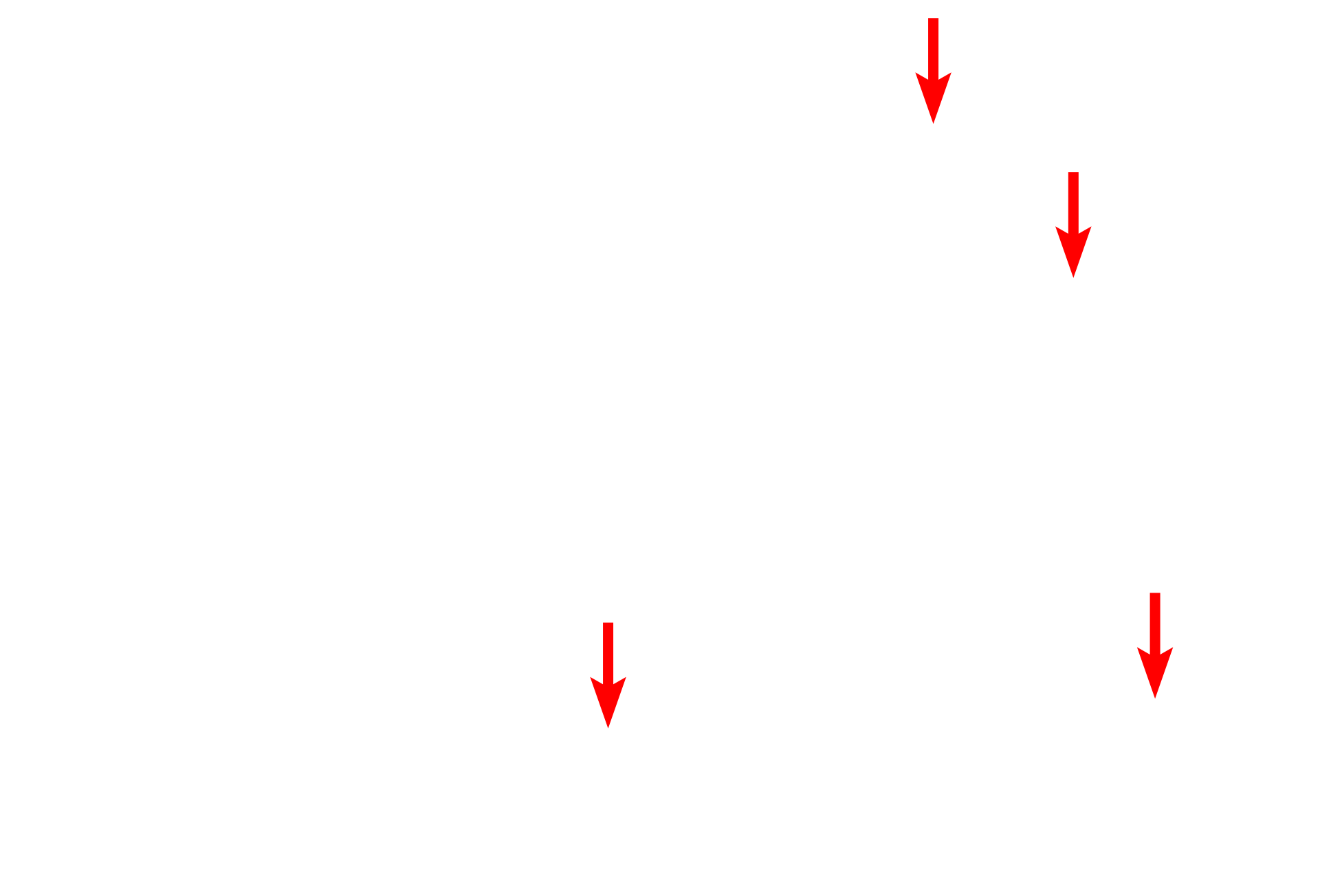
A bands >
The A band is composed of thick myofilaments overlapping a portion of the thin myofilaments.
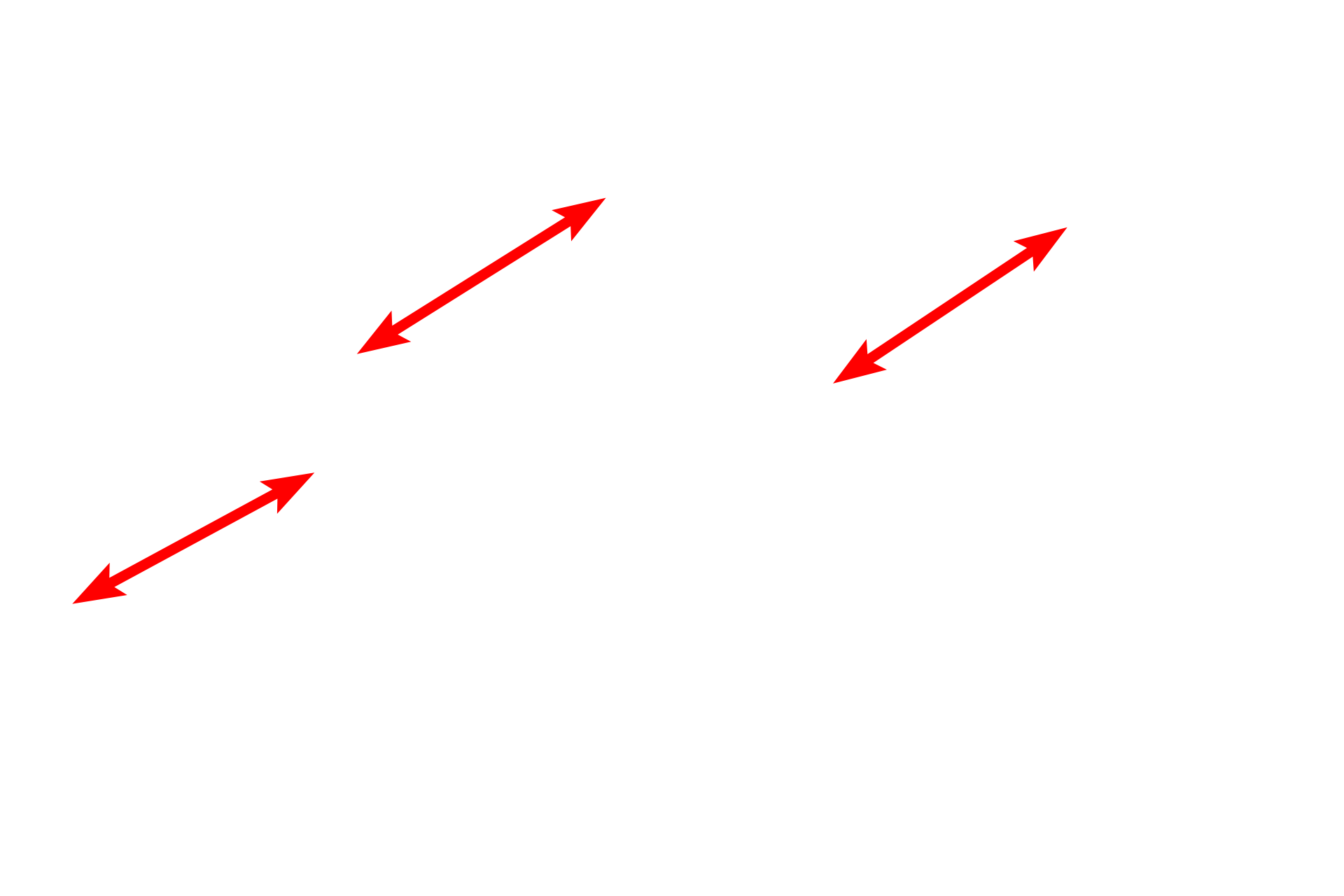
- H bands >
The H band contains thick myofilaments only. When the muscle is contracting, thin filaments slide past the thick filaments, thereby narrowing the H band.
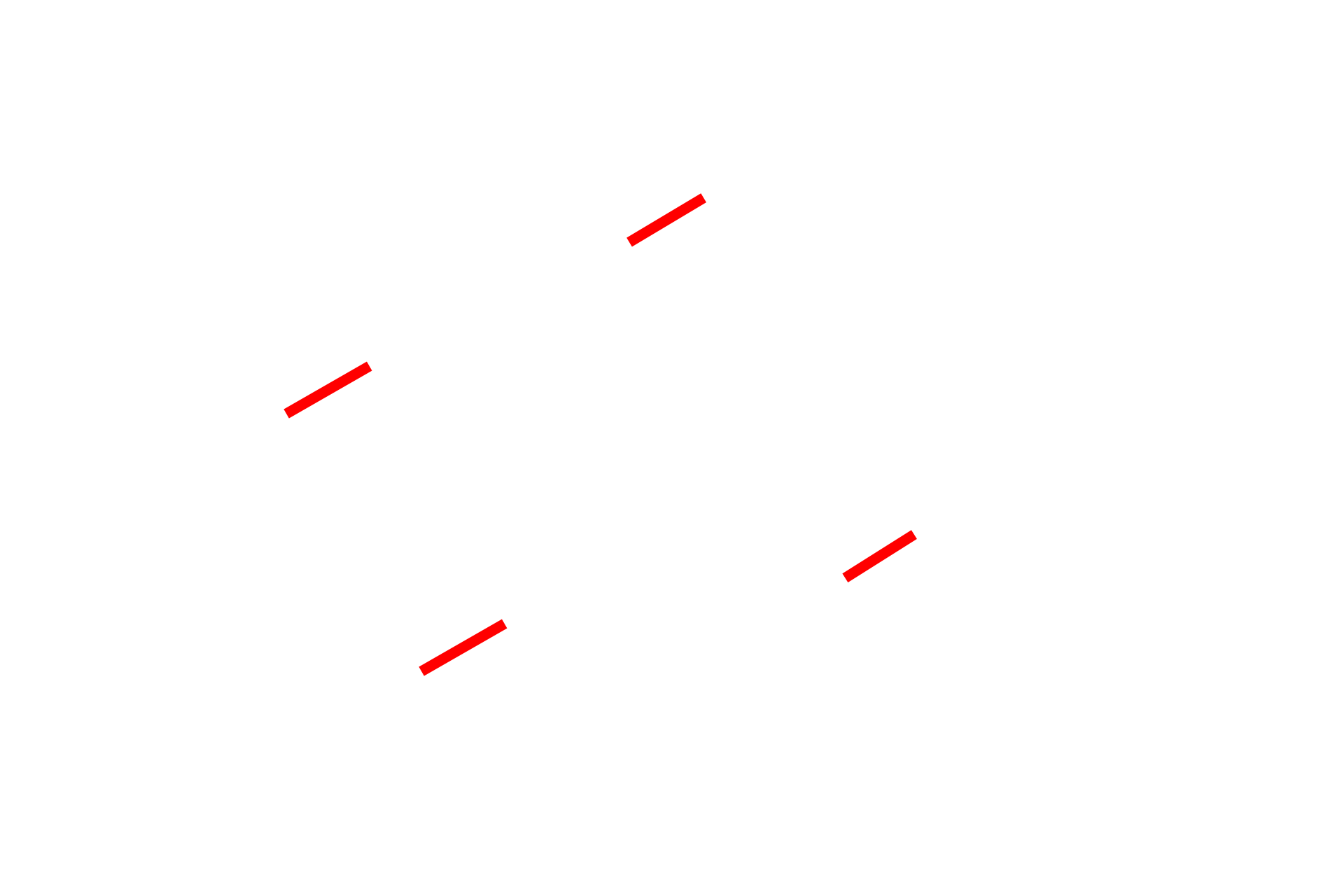
- M lines >
The M line bisects the H band and contains myosin-binding proteins that maintain the alignment of the thick myofilaments.
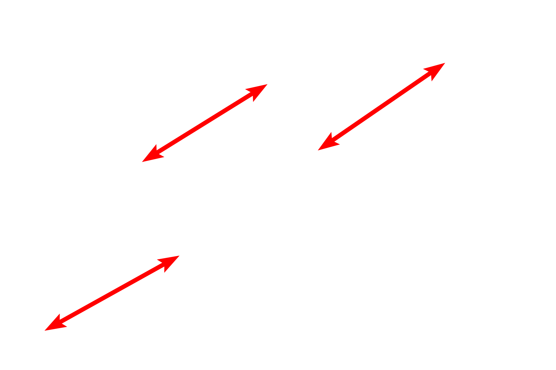
I bands >
The I band contains thin myofilaments only. When the muscle is contracting, the Z lines are brought closer together, decreasing the width of the I band.
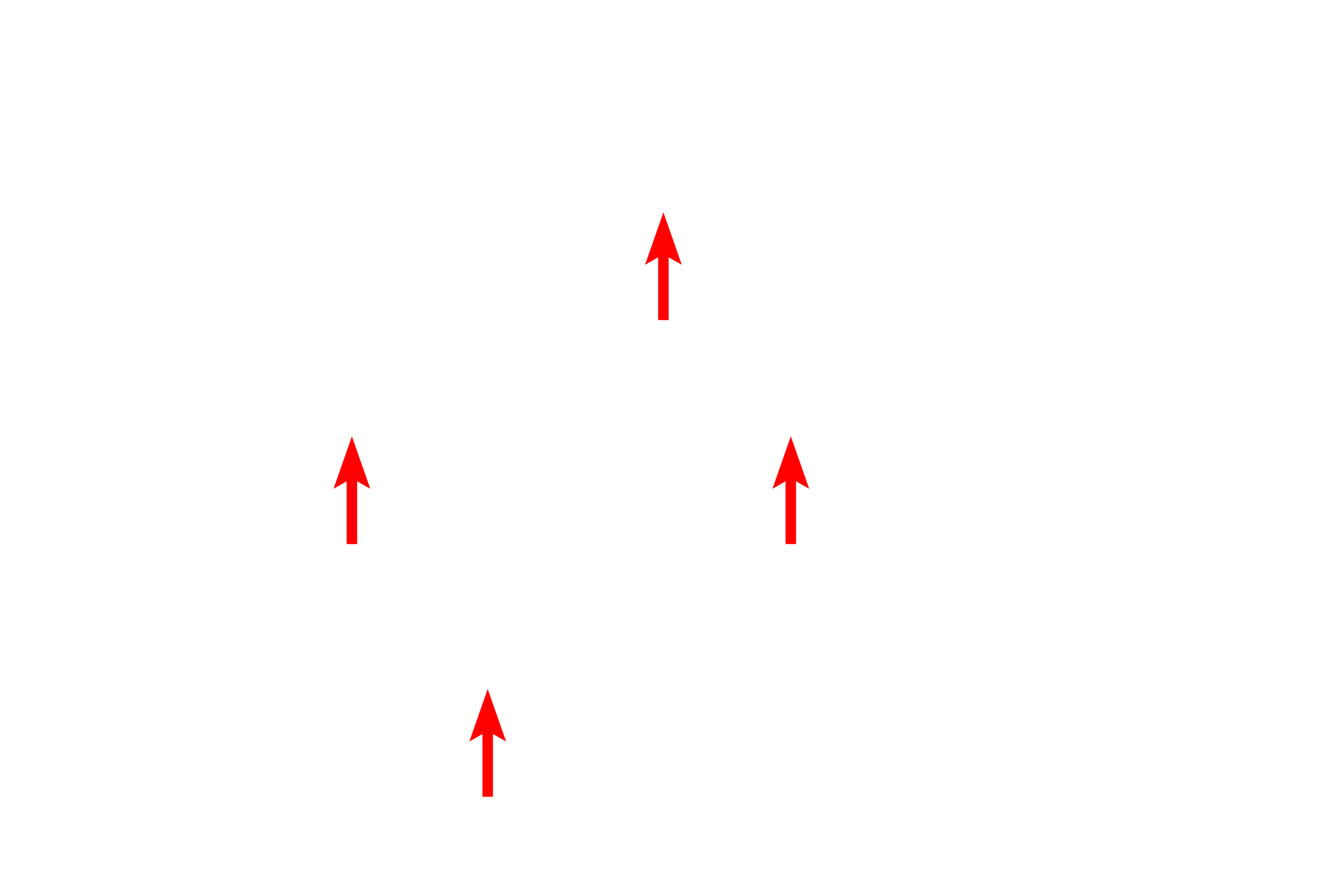
- Z lines >
The Z line bisects the I band and is the attachment site for the thin myofilaments.
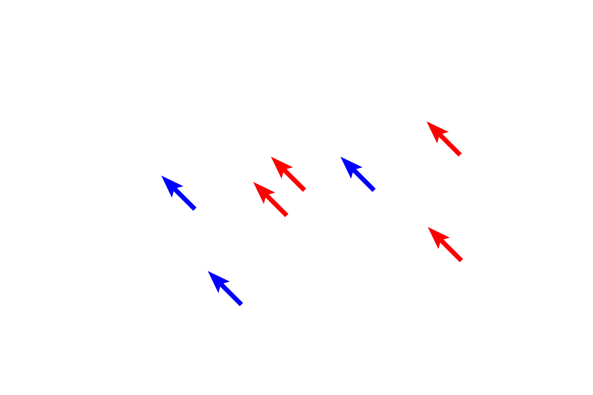
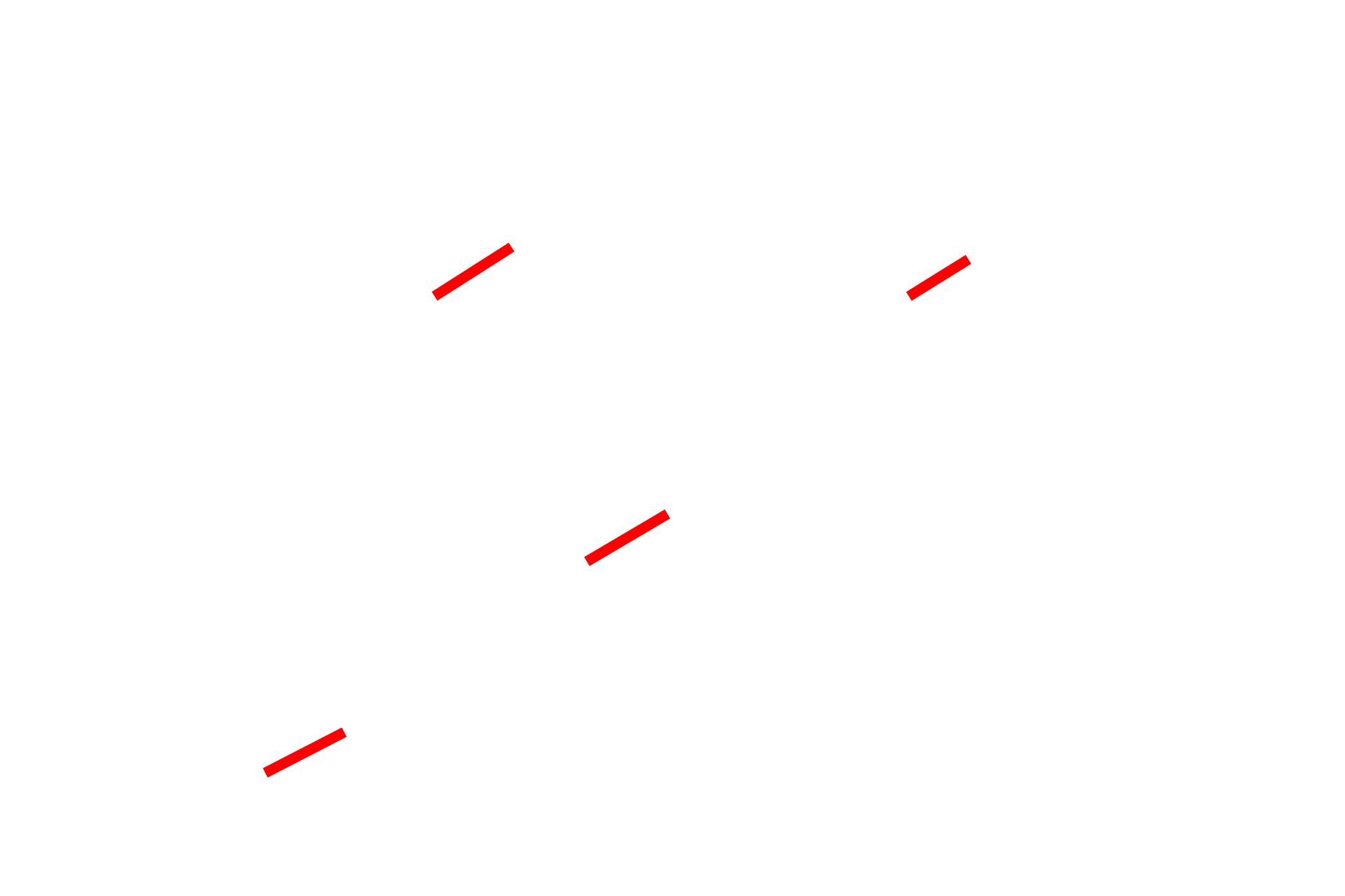
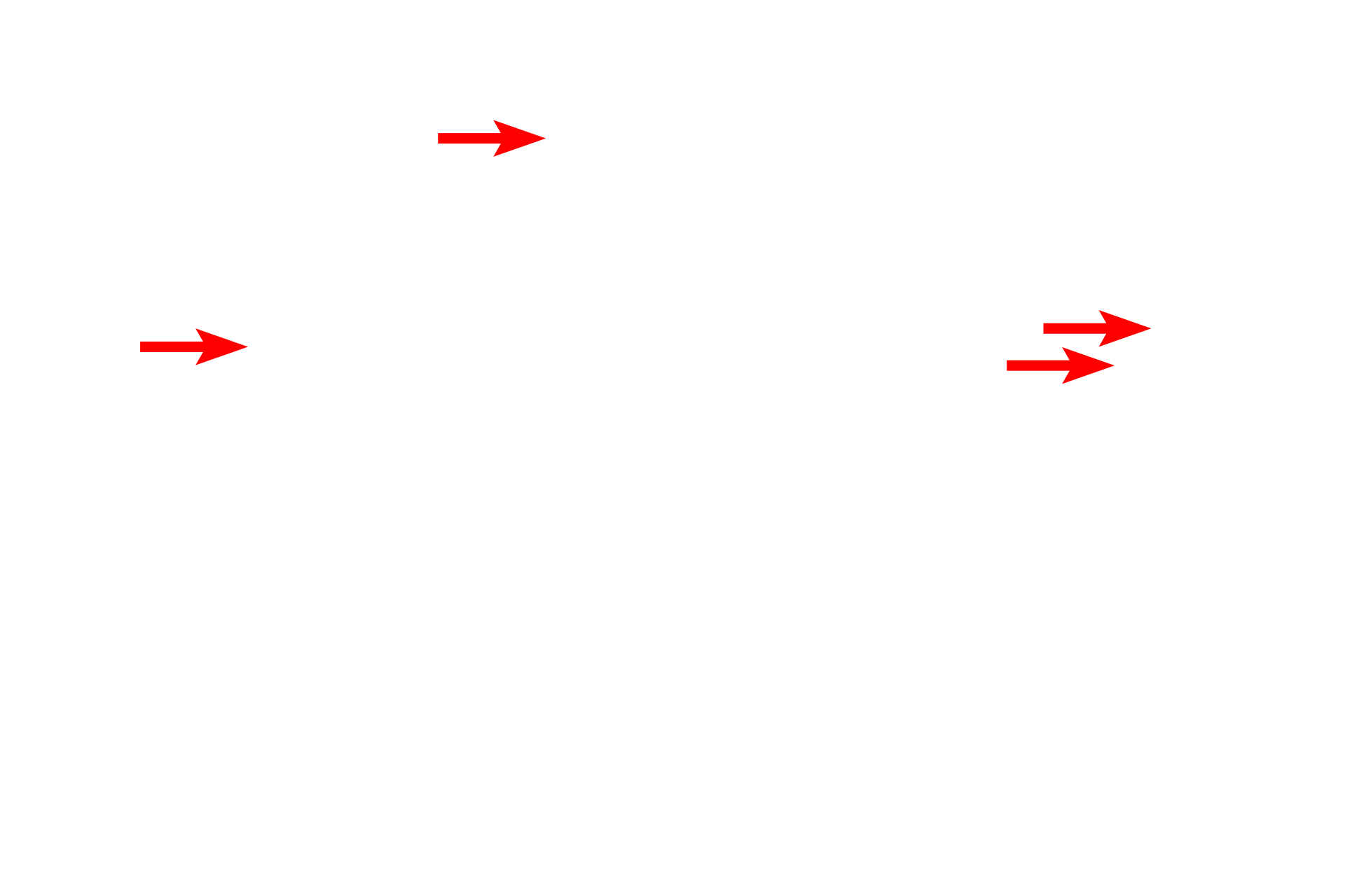
Myofilaments >
Thick myofilaments (red arrows) are broader than thin myofilaments and, therefore, are easier to identify. The thin myofilaments are difficult to resolve in this preparation, but are located in the I band (blue arrows) and extend between the thick myofilaments in the A band.
Mitochondria >
The high energy demands of cardiac muscle are reflected by the large number of mitochondria and glycogen granules.
Glycogen granules
The high energy demands of cardiac muscle are reflected by the large number of mitochondria and glycogen granules.