
Intercalated disc
This electron micrograph shows an intercalated disc attaching two adjacent cardiac muscle fibers. It is formed by the interdigitating membranes of the fibers. The junction consists of an adherent cell junction called a fascia adherens, which is similar to the zonula adherens in epithelial cells. Additionally, there are gap junctions that allow for ionic continuity between fibers, permitting waves of contraction to spread across the myocardium. The electron density is produced by the junctional proteins along with additional associated proteins. 5000x
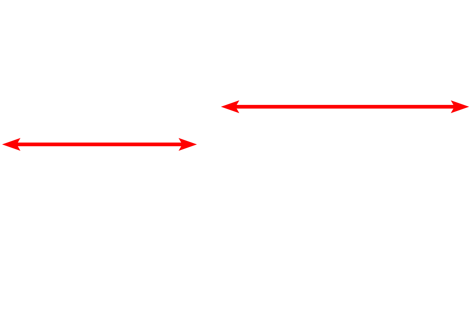
Cardiac muscle fibers
This electron micrograph shows an intercalated disc attaching two adjacent cardiac muscle fibers. It is formed by the interdigitating membranes of the fibers. The junction consists of an adherent cell junction called a fascia adherens, which is similar to the zonula adherens in epithelial cells. Additionally, there are gap junctions that allow for ionic continuity between fibers, permitting waves of contraction to spread across the myocardium. The electron density is produced by the junctional proteins along with additional associated proteins. 5000x
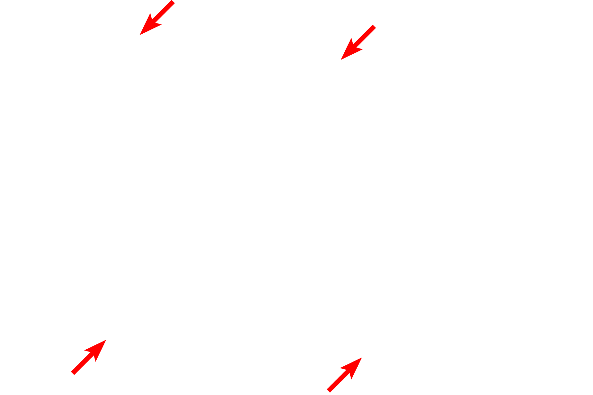
Sarcolemma
This electron micrograph shows an intercalated disc attaching two adjacent cardiac muscle fibers. It is formed by the interdigitating membranes of the fibers. The junction consists of an adherent cell junction called a fascia adherens, which is similar to the zonula adherens in epithelial cells. Additionally, there are gap junctions that allow for ionic continuity between fibers, permitting waves of contraction to spread across the myocardium. The electron density is produced by the junctional proteins along with additional associated proteins. 5000x
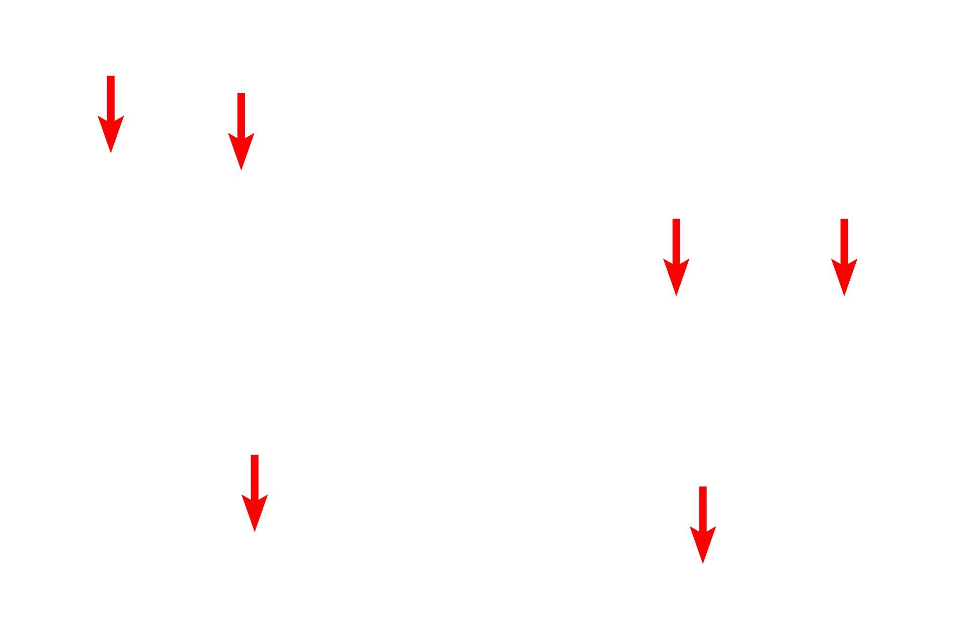
Mitochondria
Numerous mitochondria are present between the myofibrils and just beneath the sarcolemma. Mitochondria provide the large amount of energy needed for the continually-contracting fibers.
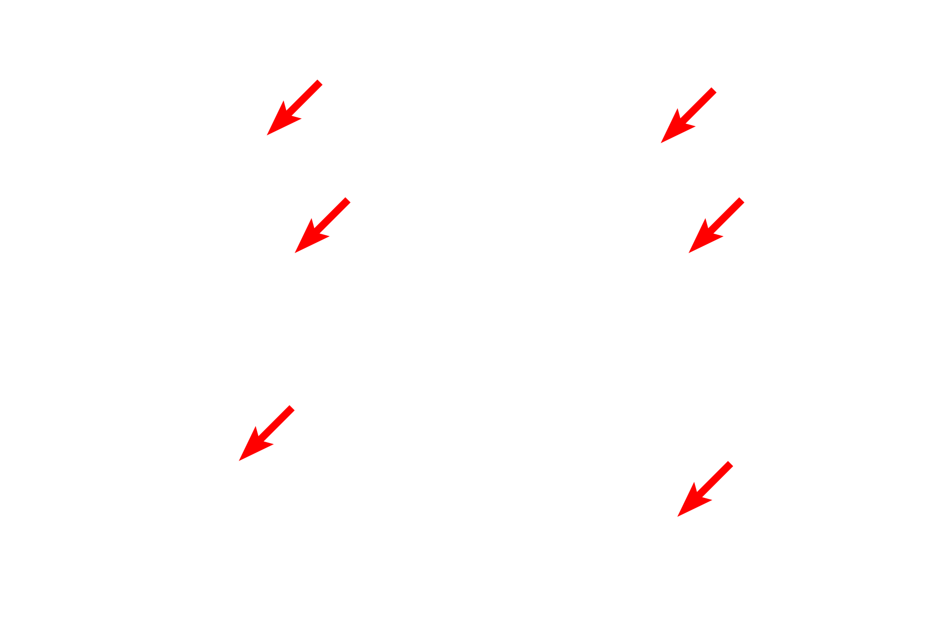
Myofibrils >
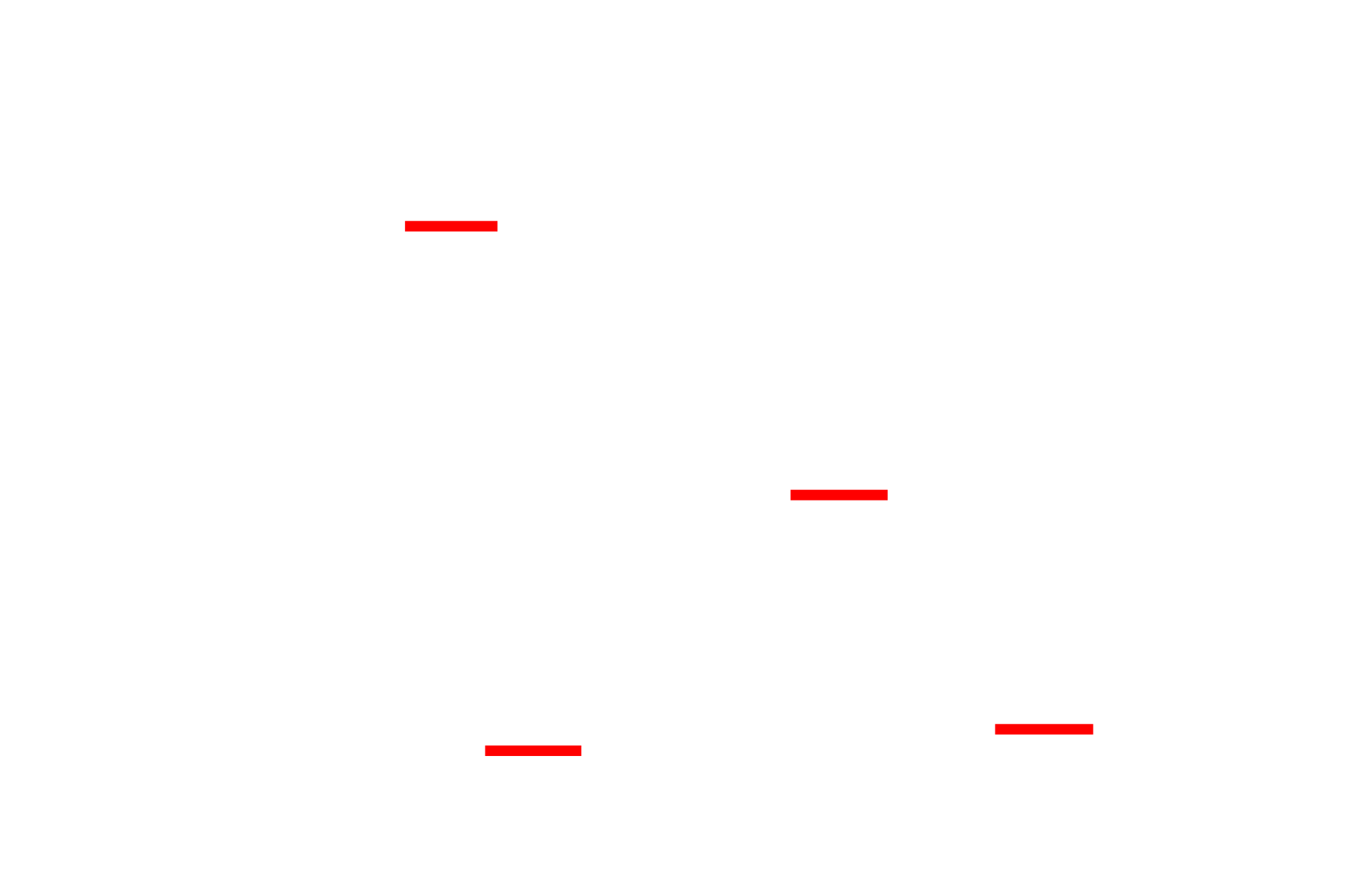
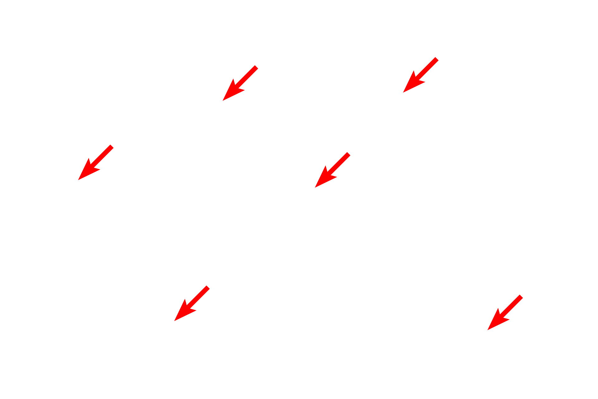
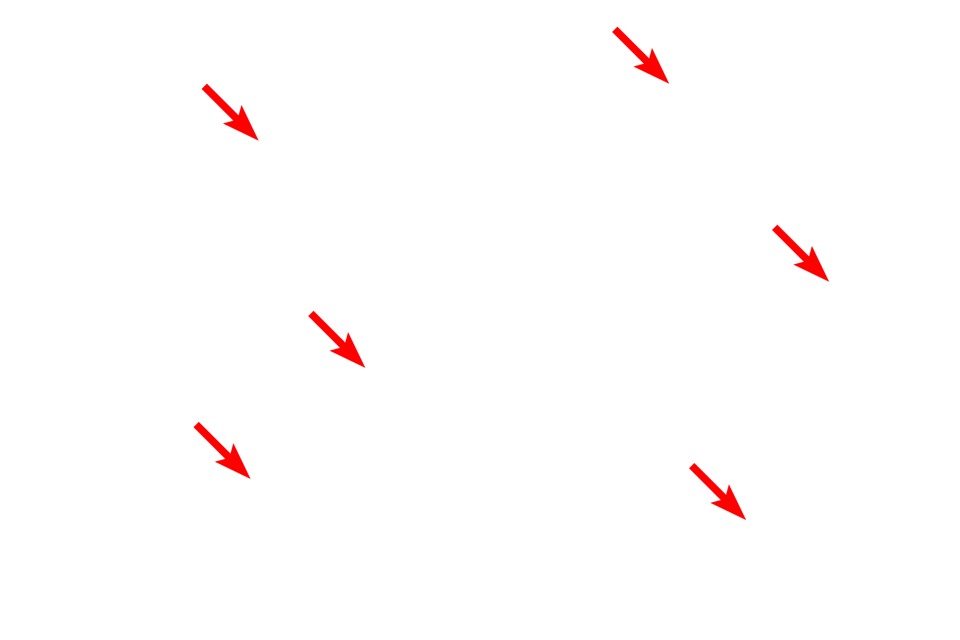
Myofibrils show the same banding pattern as that seen in skeletal muscle fibers, however they are not distinct in this micrograph. Visible here are sarcomeres, Z lines and M lines.
- Sarcomeres
Myofibrils show the same banding pattern as that seen in skeletal muscle fibers, however they are not distinct in this micrograph. Visible here are sarcomeres, Z lines and M lines.
- Z lines
Myofibrils show the same banding pattern as that seen in skeletal muscle fibers, however they are not distinct in this micrograph. Visible here are sarcomeres, Z lines and M lines.
- M lines
Myofibrils show the same banding pattern as that seen in skeletal muscle fibers, however they are not distinct in this micrograph. Visible here are sarcomeres, Z lines and M lines.
Image source >
This electron micrograph, originally published by J.A.G. Rhodin, some from the University of Michigan collection.