
Cardiac muscle
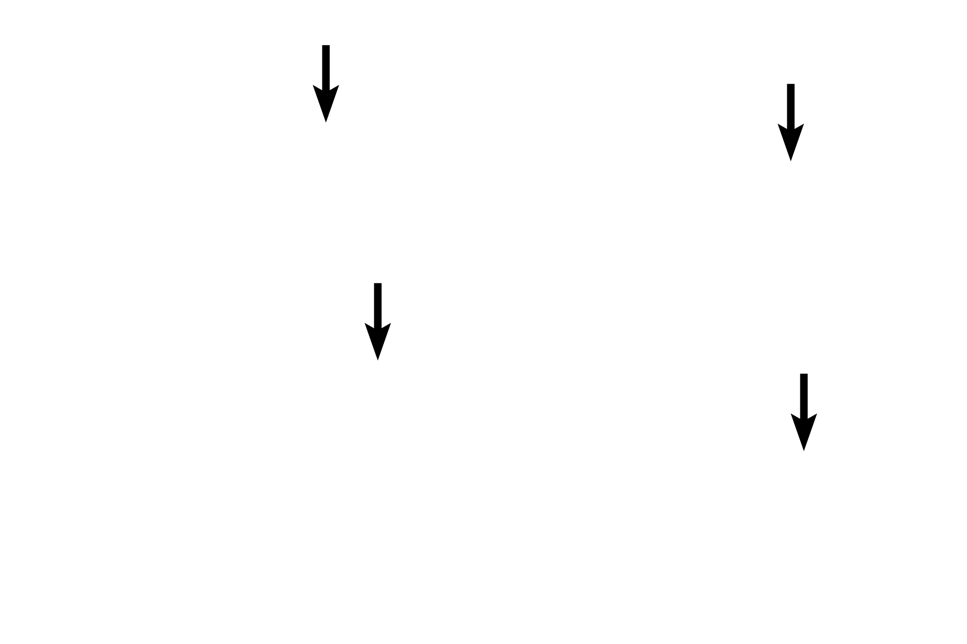
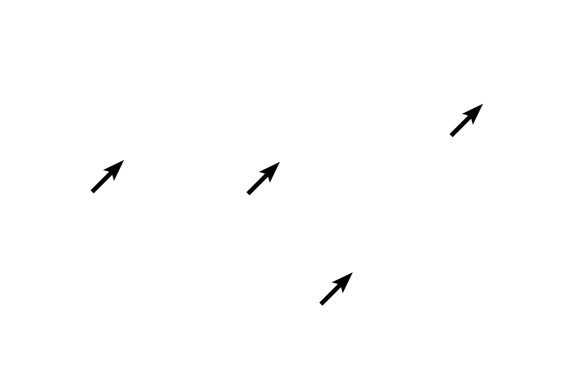
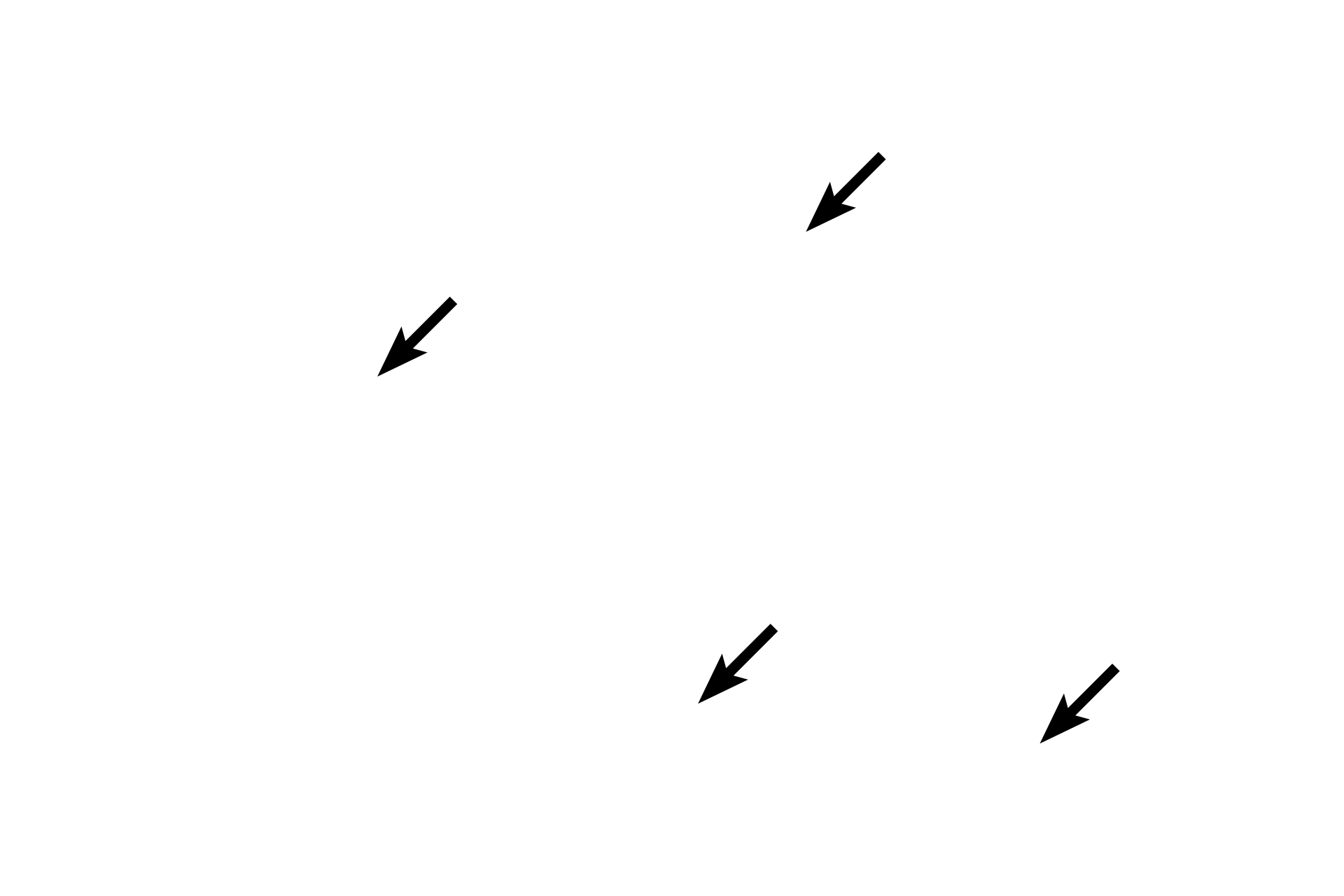
Cardiac muscle fibers are unique in that they branch and interlace. This hematoxylin and eosin stained, longitudinal section shows cardiac muscle fibers forming a feltwork in the myocardium of the heart. Cardiac muscle fibers are striated, though the cross striations are not as prominent as in skeletal muscle fibers. 400x
Muscle fibers
Cardiac muscle fibers are unique in that they branch and interlace. This hematoxylin and eosin stained, longitudinal section shows cardiac muscle fibers forming a feltwork in the myocardium of the heart. Cardiac muscle fibers are striated, though the cross striations are not as prominent as in skeletal muscle fibers. 400x

Fiber branching
Cardiac muscle fibers are unique in that they branch and interlace. This hematoxylin and eosin stained, longitudinal section shows cardiac muscle fibers forming a feltwork in the myocardium of the heart. Cardiac muscle fibers are striated, though the cross striations are not as prominent as in skeletal muscle fibers. 400x
Nuclei
Cardiac muscle fibers are unique in that they branch and interlace. This hematoxylin and eosin stained, longitudinal section shows cardiac muscle fibers forming a feltwork in the myocardium of the heart. Cardiac muscle fibers are striated, though the cross striations are not as prominent as in skeletal muscle fibers. 400x

Striations
Cardiac muscle fibers are unique in that they branch and interlace. This hematoxylin and eosin stained, longitudinal section shows cardiac muscle fibers forming a feltwork in the myocardium of the heart. Cardiac muscle fibers are striated, though the cross striations are not as prominent as in skeletal muscle fibers. 400x
Intercalated discs
Cardiac muscle fibers are unique in that they branch and interlace. This hematoxylin and eosin stained, longitudinal section shows cardiac muscle fibers forming a feltwork in the myocardium of the heart. Cardiac muscle fibers are striated, though the cross striations are not as prominent as in skeletal muscle fibers. 400x
Lipofuscin pigment >
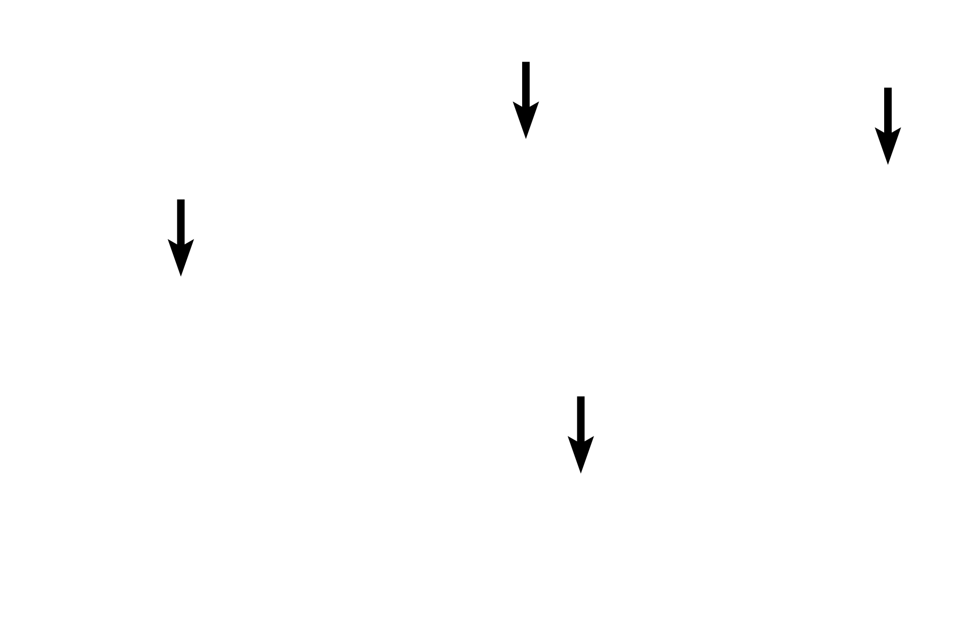
Lipofuscin is a pigment residue of lysosomal digestion and appears as fine, membrane-bound yellowish-brown granules in the cytoplasm. Lipofuscin is composed of oxidized lipids and proteins that accumulate in cells during normal aging. Lipofuscin is particularly abundant in long-lived cells such as these muscle fibers and neurons.
Image source >
This image is owned by Dr. Jose Luis Calvo and is used under a royalty-free agreement.
 PREVIOUS