
Overview of surface epithelia
Surface epithelia cover the exterior of the body and line all interior surfaces. These epithelia are classified by the shape of the cells immediately facing the surface or lumen and by the number of cell layers composing the epithelium. Most epithelia show polarity, are avascular and rest on a basement membrane that they produce with the underlying connective tissue.

Simple epithelia >
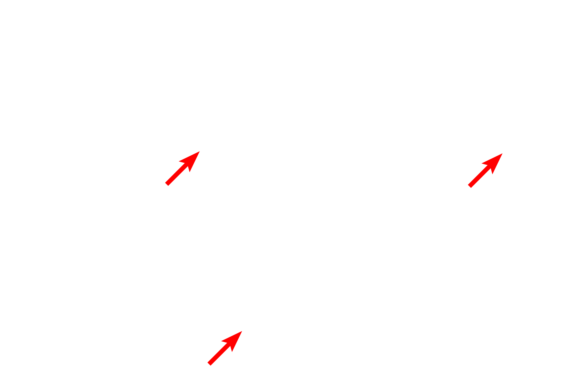
Simple epithelia consist of a single layer of cells that all lie on the basement membrane (arrows). The cells may be squamous shaped (like a fried egg), cuboidal, columnar, or a combination of cell shapes. Only simple epithelia have surface modifications such as microvilli and cilia.

- Squamous >
Simple squamous epithelium consists of a single layer of flattened cells with very thin cytoplasm, resembling the white of a fried egg. The nucleus of the squamous cell often bulges into the lumen, resembling the egg yolk. Because of its thinness, simple squamous epithelium lines structures where rapid transport is required, such as capillaries and lung alveoli.

- Cuboidal >
Simple cuboidal epithelium consists of a single layer of cube-shaped cells. Simple cuboidal epithelia line small ducts of glands and many of the tubules in the kidney.

- Columnar >
Simple columnar epithelium is composed of a single layer of column-shaped cells. The oval nuclei in this epithelium are usually located in the basal half to the center of each cell. Simple columnar epithelium may possess microvilli, such as in the small intestine, where they aid in absorption of digested food, or cilia, such as in the oviduct where they provide transport for germ cells.

- Pseudostratified >
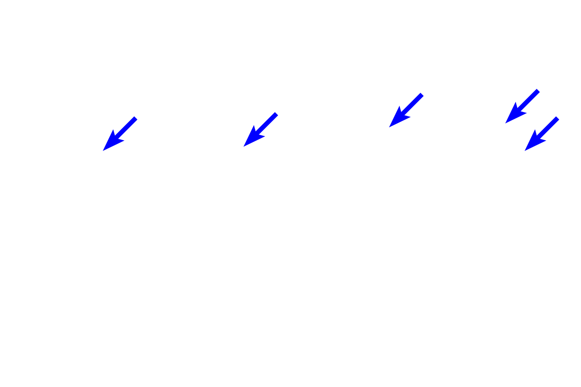
Pseudostratified epithelium is really a simple epithelium because each cell rests on the basement membrane. However, not all cells extend to the lumen, giving this tissue the appearance of being stratified because nuclei appear at different levels. Pseudostratified epithelium lines many of the respiratory passageways where it possesses cilia (arrow), an indication that this is truly a simple epithelium.

Stratified epithelia >
Stratified epithelia are composed of two or more cell layers and are classified by the shape of the surface layer of cells. Stratified epithelia are not involved with transport or absorption, but rather, they provide a protective layer for to the underlying tissues. The basal layer is mitotically active to provide new cells for the epithelium. Stratified epithelial do not have surface specializations.

- Squamous moist >
Stratified squamous moist (or nonkeratinized) epithelium consists of multiple cell layers. The uppermost layers of cells are squamous in shape and are living, thus providing a moist surface. The basal layer of cells is mitotically active, generating new cells for the epithelium. This epithelium lines interior body surface such as the esophagus and vagina, that require protection from abrasion.

- Squamous dry >
Stratified squamous dry (or keratinized) epithelium is composed of multiple cell layers that flatten as they approach the surface. These epithelial cells produce a cornified layer at the surface (arrow) that is composed of flattened, non-nucleated, dead cells filled with the protein keratin. Stratified squamous keratinized epithelium covers the surface of the body forming the outer layer of the skin.

- Columnar >
Stratified columnar epithelium is composed of multiple layers, the uppermost of which is column-shaped. This epithelium is not found in a definitive location but is frequently seen in areas of epithelial transition, for example, in ducts as their linings are modified from a simple epithelium to a stratified epithelium.

- Transitional >
Transitional epithelium is found in the urinary system where it allows for distension of organs, e.g., the urinary bladder. This epithelium is also classified as stratified cuboidal because its surface cells frequently assume a cuboidal shape when the organ is not distended. These surface cells are called dome cells (arrow) because of their bulging shape. However, they flatten to a squamous shape when the epithelium is stretched, thus the name “transitional.”
Basement membrane >
All epithelia rest on a basement membrane, which consists of a basal lamina, produced by the epithelial cells, and a reticular lamina, produced by fibroblasts in the underlying connective tissue. Because the basal lamina and reticular lamina cannot be resolved with the light microscope, the term basement membrane is used in light microscopy to refer to both of these structures, collectively.
Nuclear shapes >
The shape of the nucleus is a good indicator of the overall shape of the cell. In squamous cells, the nucleus is flat, in cuboidal cells the nucleus is spherical, and in columnar cells the nucleus is oval. These features are helpful in studying tissue sections where cell outlines are not always readily visible.