
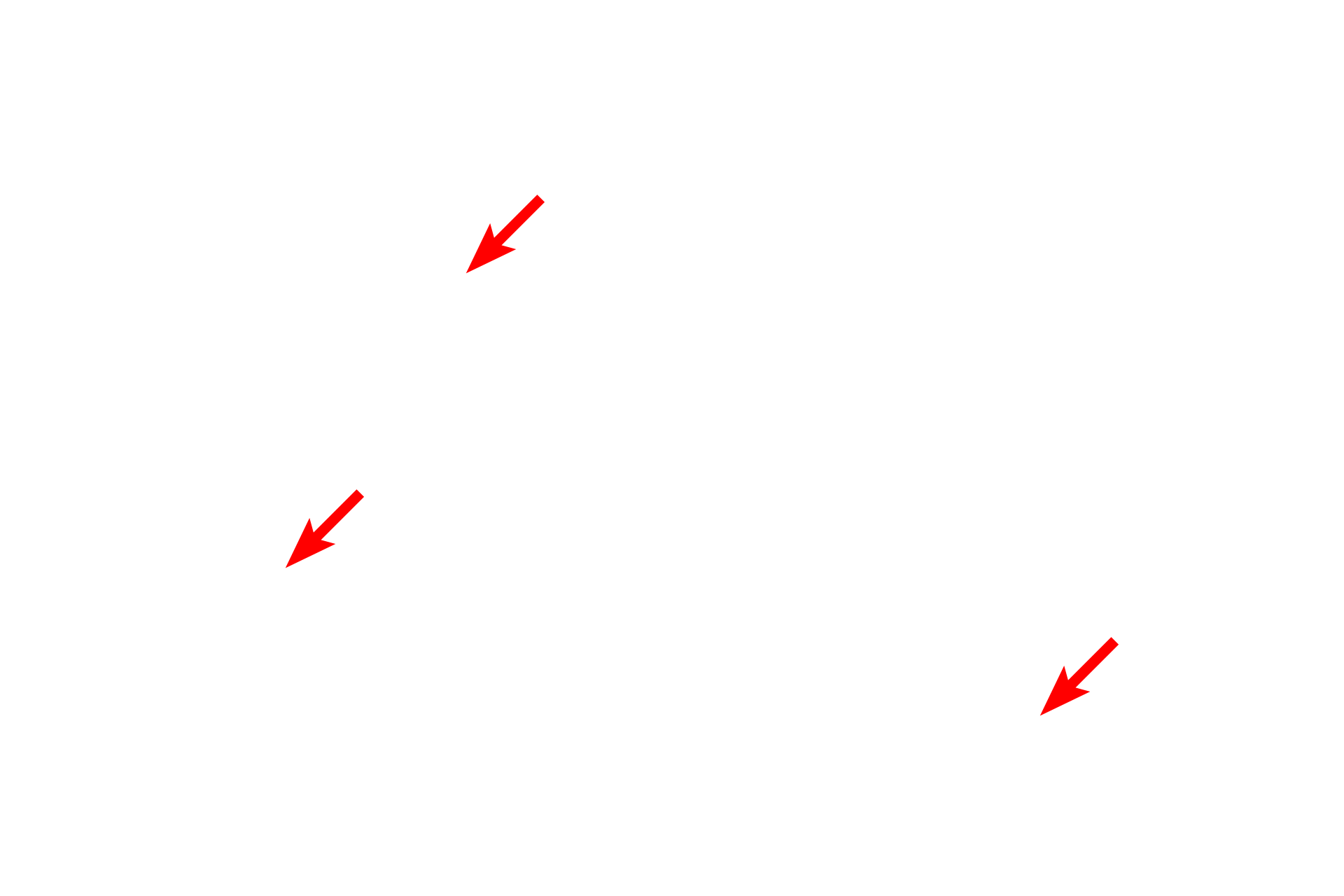
Reticular connective tissue
Reticular fibers are composed of Type III collagen and are unique from Type I collagen fibers in that they branch. Additionally, reticular fibers are glycosylated. They require special stains to be clearly identified, such as silver (seen here) or periodic acid Schiff (PAS). Lymph node 1000x
Reticular fibers
Reticular fibers are composed of Type III collagen and are unique from Type I collagen fibers in that they branch. Additionally, reticular fibers are glycosylated. They require special stains to be clearly identified, such as silver (seen here) or periodic acid Schiff (PAS). Lymph node 1000x
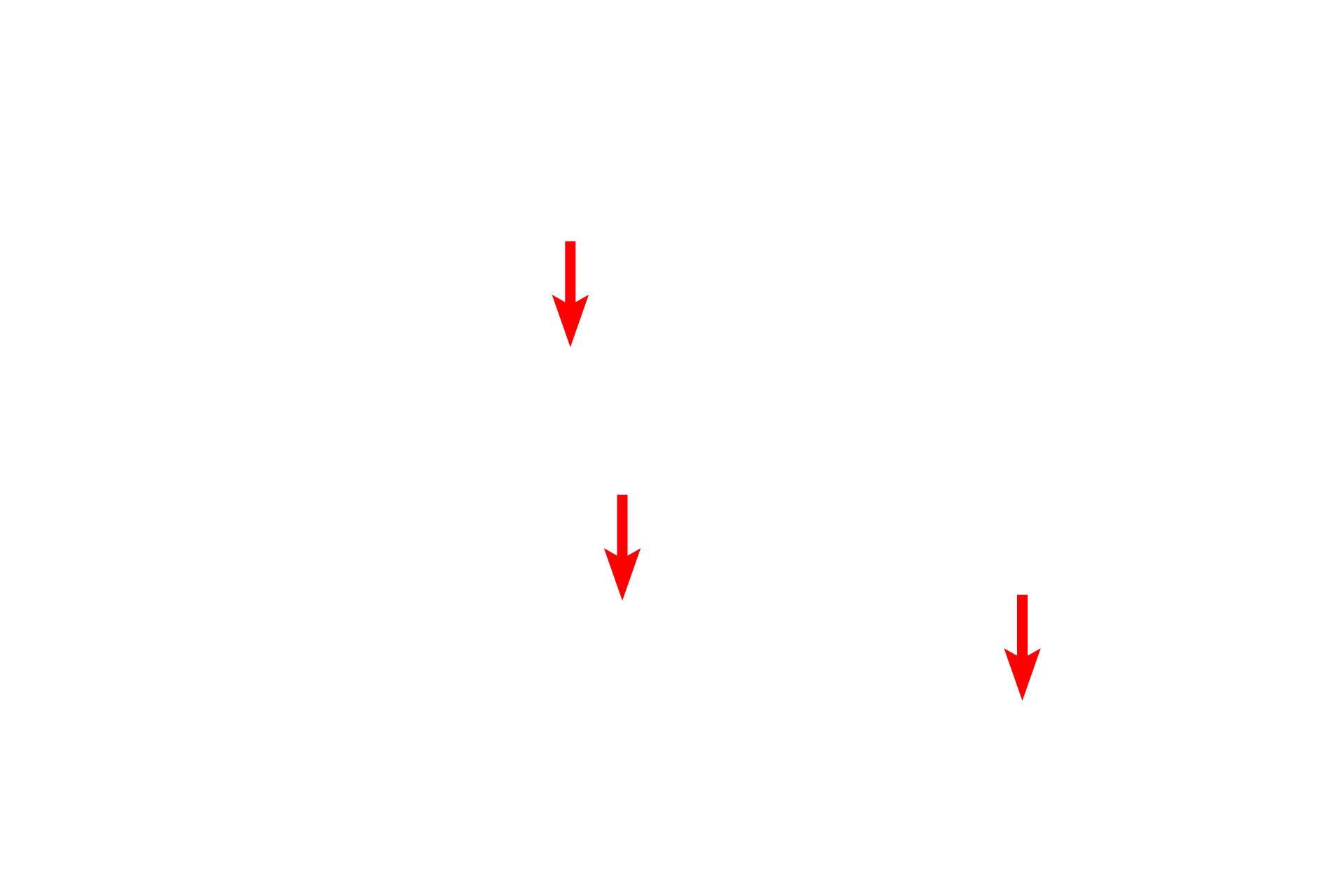
Branching points
Reticular fibers are composed of Type III collagen and are unique from Type I collagen fibers in that they branch. Additionally, reticular fibers are glycosylated. They require special stains to be clearly identified, such as silver (seen here) or periodic acid Schiff (PAS). Lymph node 1000x