
Eosinophil
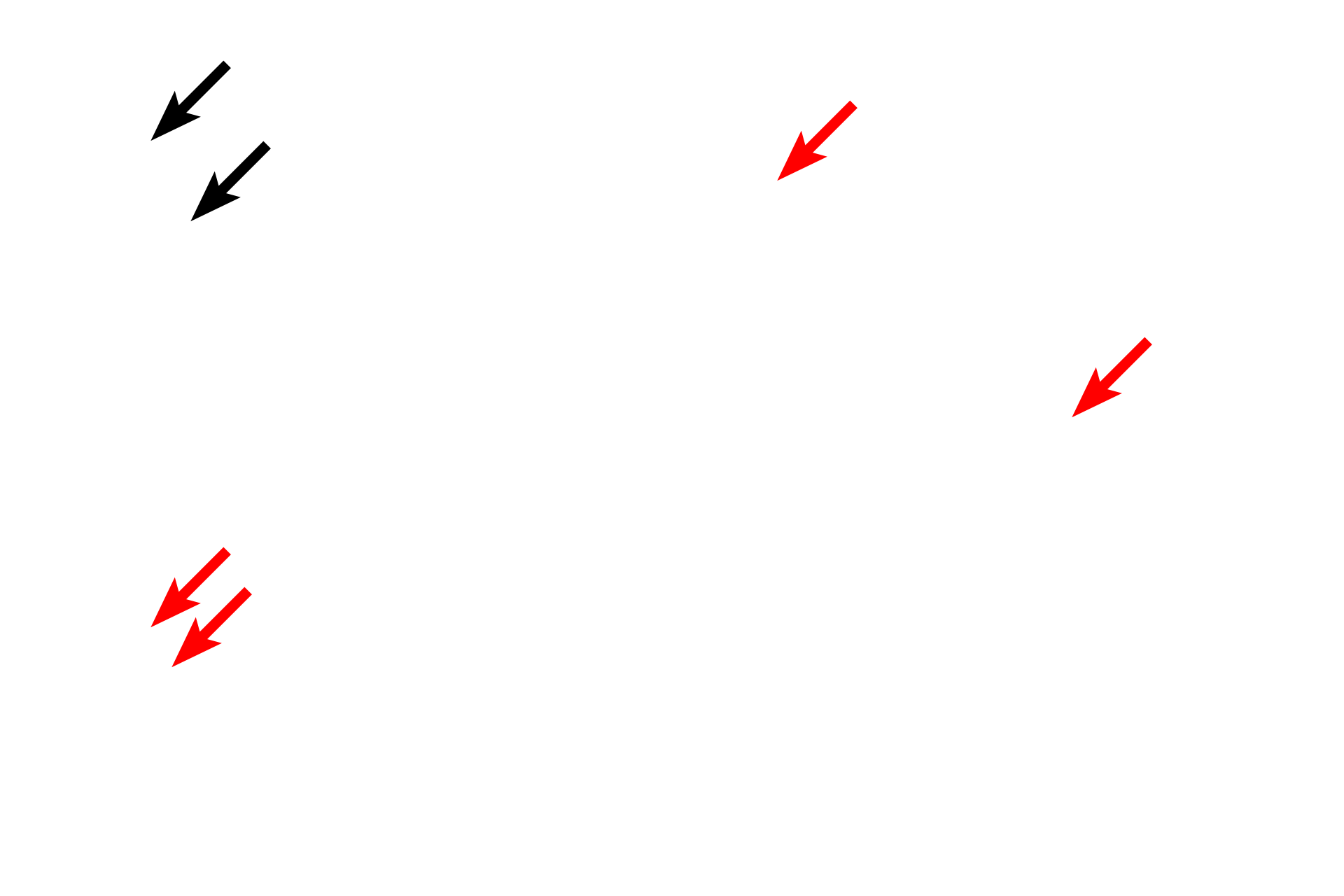
These micrographs show the cellular components of eosinophils. Note the bilobed nucleus with lobes connected by a thin region of chromatin. Like all granular leucocytes, eosinophils have both azurophilic (primary) granules and specific (secondary) granules. In eosinophils, the specific granules contain a large crystalloid body surrounded by an electron dense matrix. These granules stain intensely with eosin. The cytoplasm contains few other membranous organelles. 1000x, 10,000x
Bilobed nucleus
These micrographs show the cellular components of eosinophils. Note the bilobed nucleus with lobes connected by a thin region of chromatin. Like all granular leucocytes, eosinophils have both azurophilic (primary) granules and specific (secondary) granules. In eosinophils, the specific granules contain a large crystalloid body surrounded by an electron dense matrix. These granules stain intensely with eosin. The cytoplasm contains few other membranous organelles. 1000x, 10,000x
Specific granules >
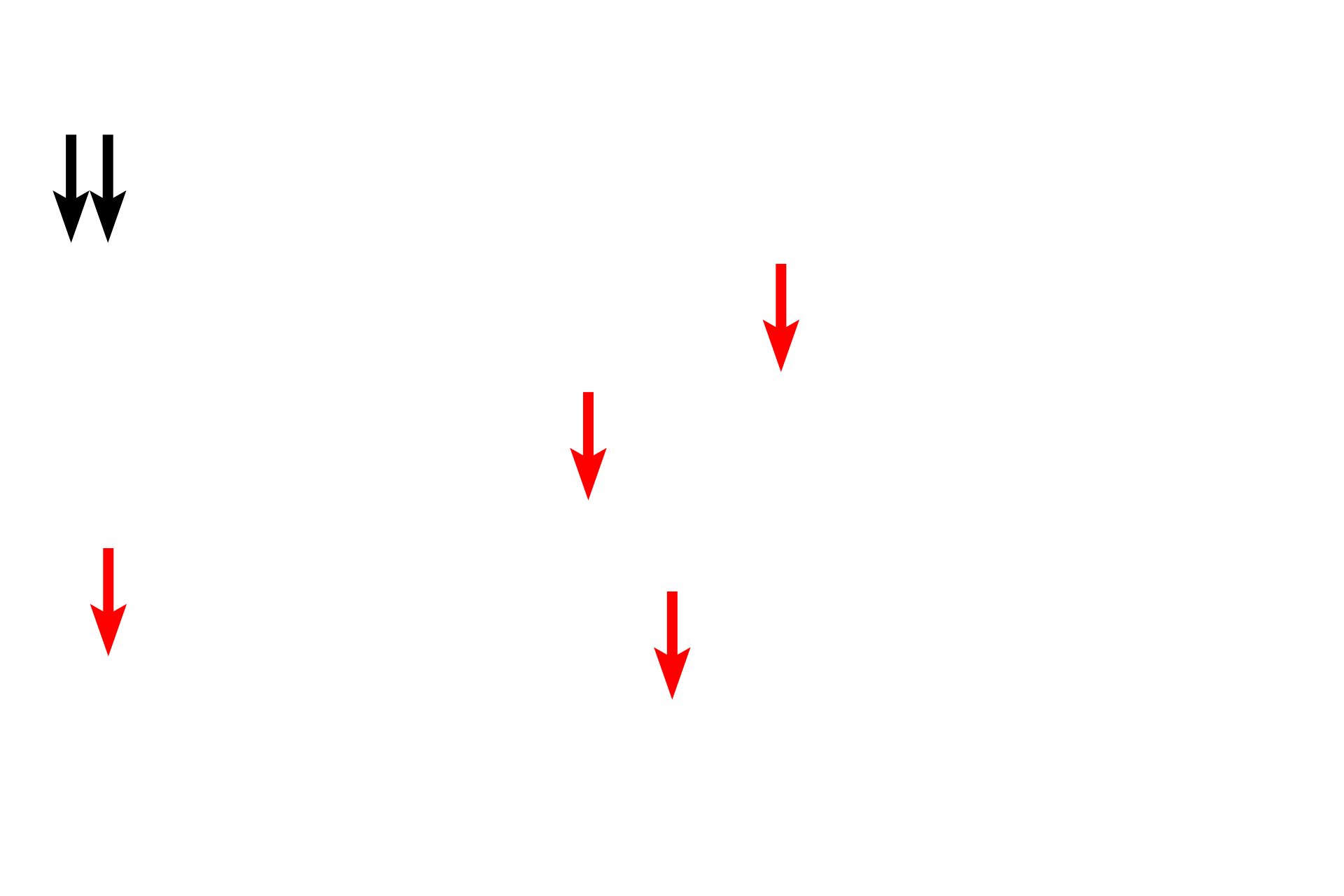
The contents of the specific granules of eosinophils have powerful cytotoxic effects on protozoan and helminthic parasites. These granules stain intensely with eosin and contain a crystalloid body that is responsible for the refractivity of the granules seen by light microscopy.
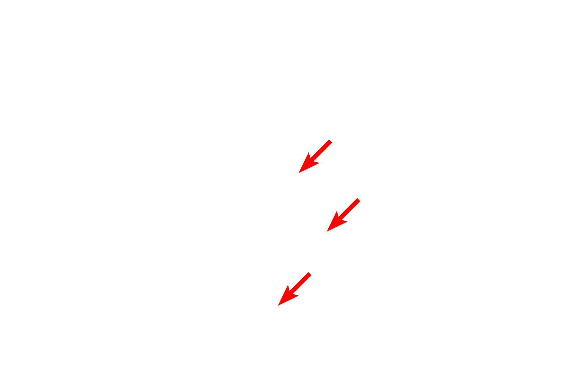
- Crystalloid bodies
The contents of the specific granules of eosinophils have powerful cytotoxic effects on protozoan and helminthic parasites. These granules stain intensely with eosin and contain a crystalloid body that is responsible for the refractivity of the granules seen by light microscopy.
Image credit >
The electron micrograph was generated by Dr. Johannes A. G. Rhodin, “An Atlas of Histology” (Oxford Press, 1974) and maintained in the University of Michigan collection.