
Mast cell
This electron micrograph shows a mast cell in loose connective tissue. The centrally located nucleus is surrounded by numerous secretory granules that contain histamine, heparin and other chemical mediators of the immune response including enzyme proteases. Bundles of collagen fibrils are also visible. 8,000x

Nucleus
This electron micrograph shows a mast cell in loose connective tissue. The centrally located nucleus is surrounded by numerous secretory granules that contain histamine, heparin and other chemical mediators of the immune response including enzyme proteases. Bundles of collagen fibrils are also visible. 8,000x
Secretory granules
This electron micrograph shows a mast cell in loose connective tissue. The centrally located nucleus is surrounded by numerous secretory granules that contain histamine, heparin and other chemical mediators of the immune response including enzyme proteases. Bundles of collagen fibrils are also visible. 8,000x

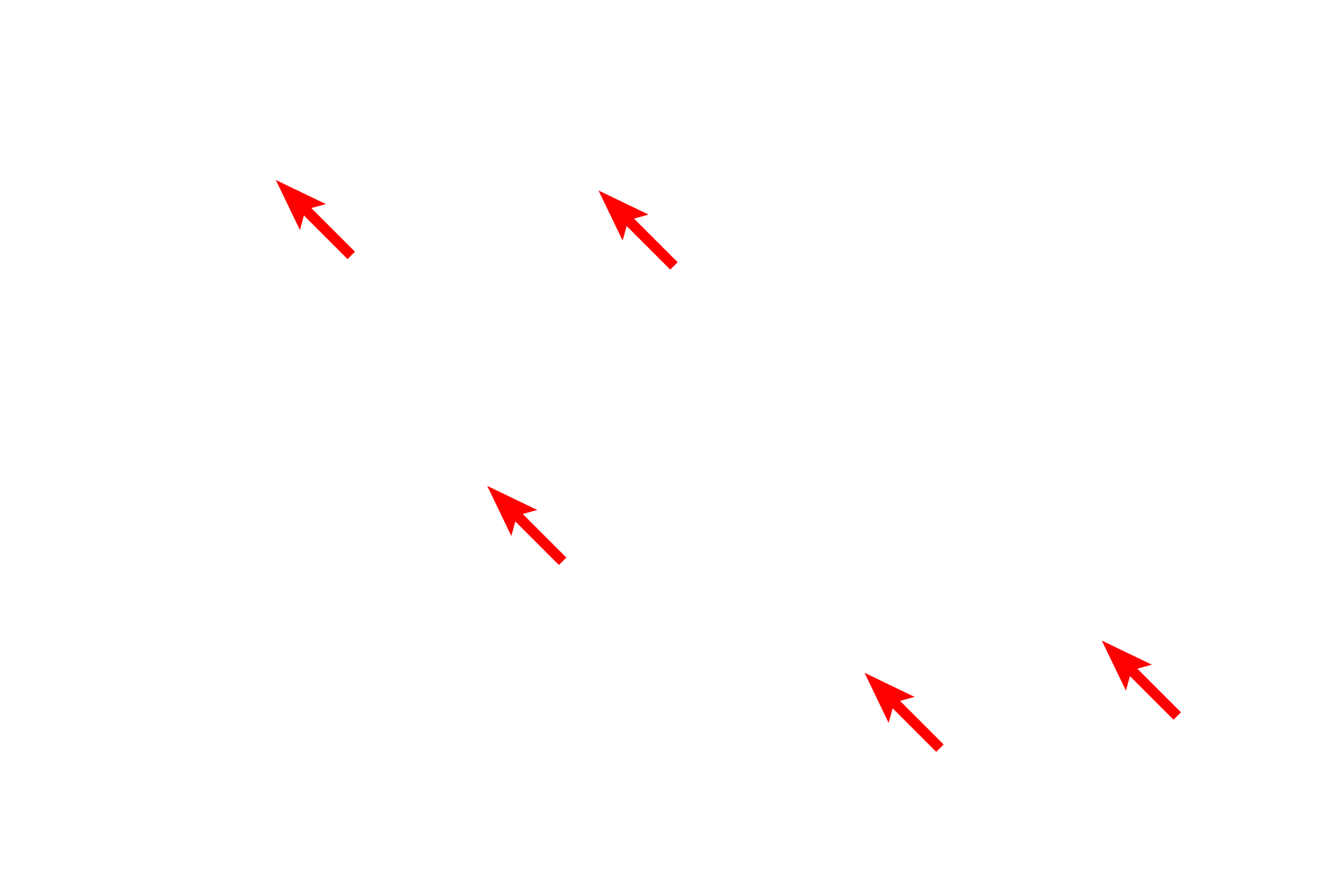
Collagen fibrils
This electron micrograph shows a mast cell in loose connective tissue. The centrally located nucleus is surrounded by numerous secretory granules that contain histamine, heparin and other chemical mediators of the immune response including enzyme proteases. Bundles of collagen fibrils are also visible. 8,000x
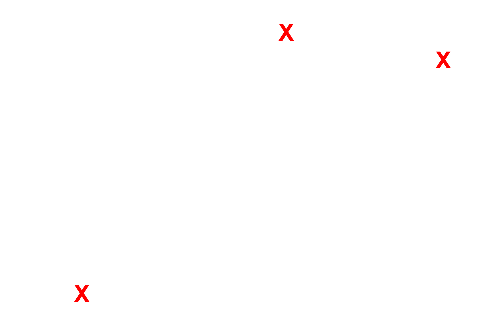
Ground substance
This electron micrograph shows a mast cell in loose connective tissue. The centrally located nucleus is surrounded by numerous secretory granules that contain histamine, heparin and other chemical mediators of the immune response including enzyme proteases. Bundles of collagen fibrils are also visible. 8,000x