
Elastic cartilage
These images compare the appearance of elastic cartilage stained with hematoxylin and eosin (left) and hematoxylin and eosin plus orcein for elastic fibers on the right. 400x
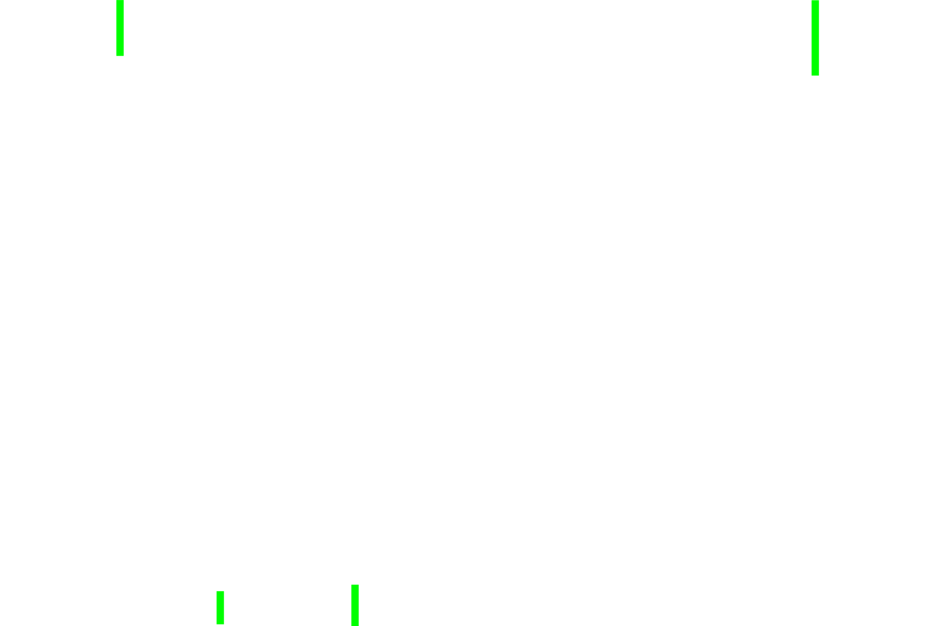
Perichondrium: fibrous layer
These images compare the appearance of elastic cartilage stained with hematoxylin and eosin (left) and hematoxylin and eosin plus orcein for elastic fibers on the right. 400x
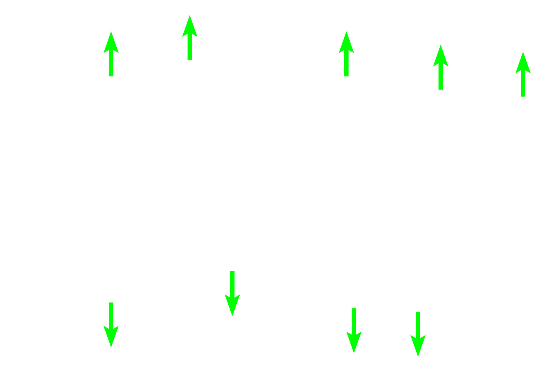
Perichondrium: chondrogenic layer
These images compare the appearance of elastic cartilage stained with hematoxylin and eosin (left) and hematoxylin and eosin plus orcein for elastic fibers on the right. 400x

Elastic cartilage
These images compare the appearance of elastic cartilage stained with hematoxylin and eosin (left) and hematoxylin and eosin plus orcein for elastic fibers on the right. 400x
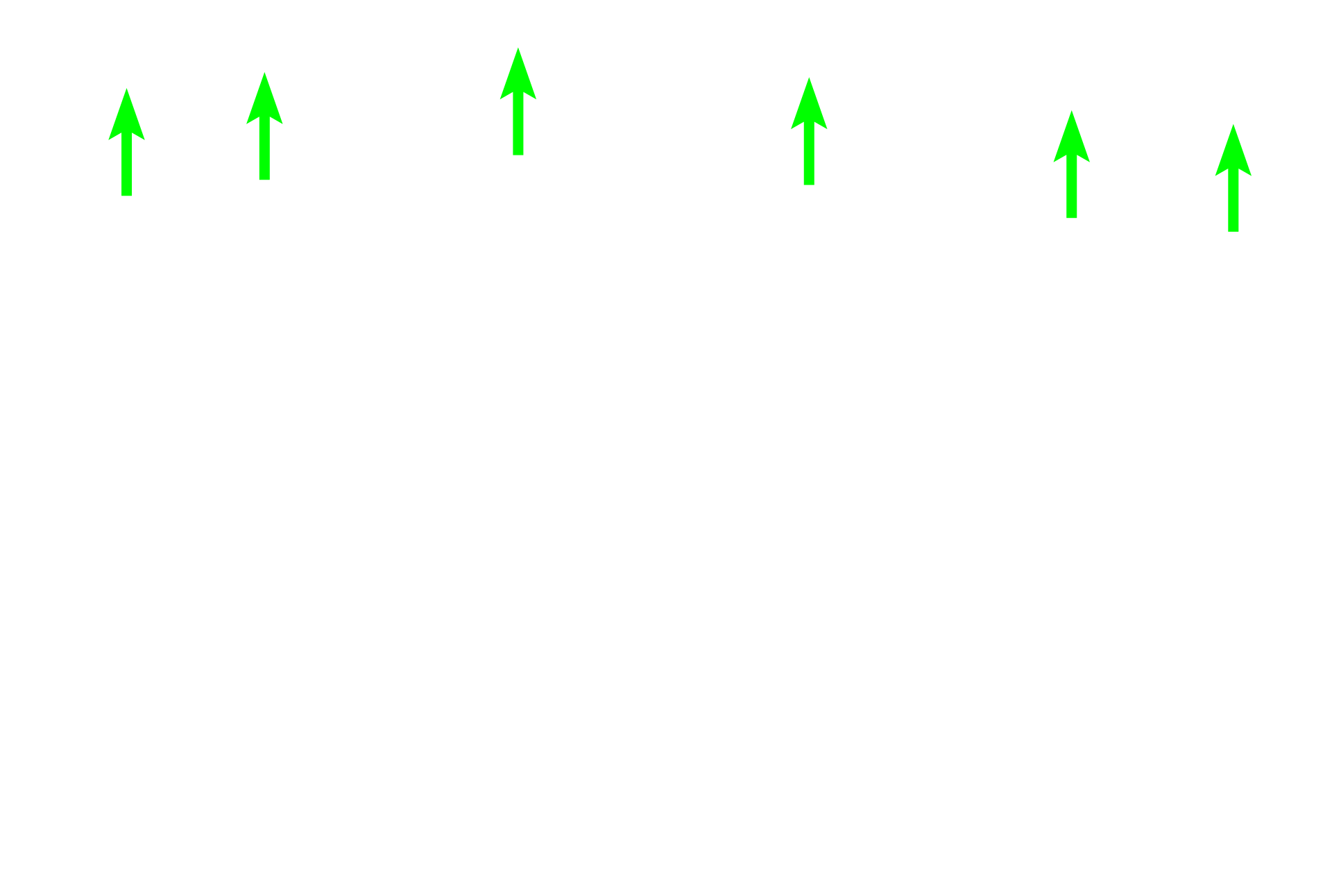
Chondroblasts
These images compare the appearance of elastic cartilage stained with hematoxylin and eosin (left) and hematoxylin and eosin plus orcein for elastic fibers on the right. 400x
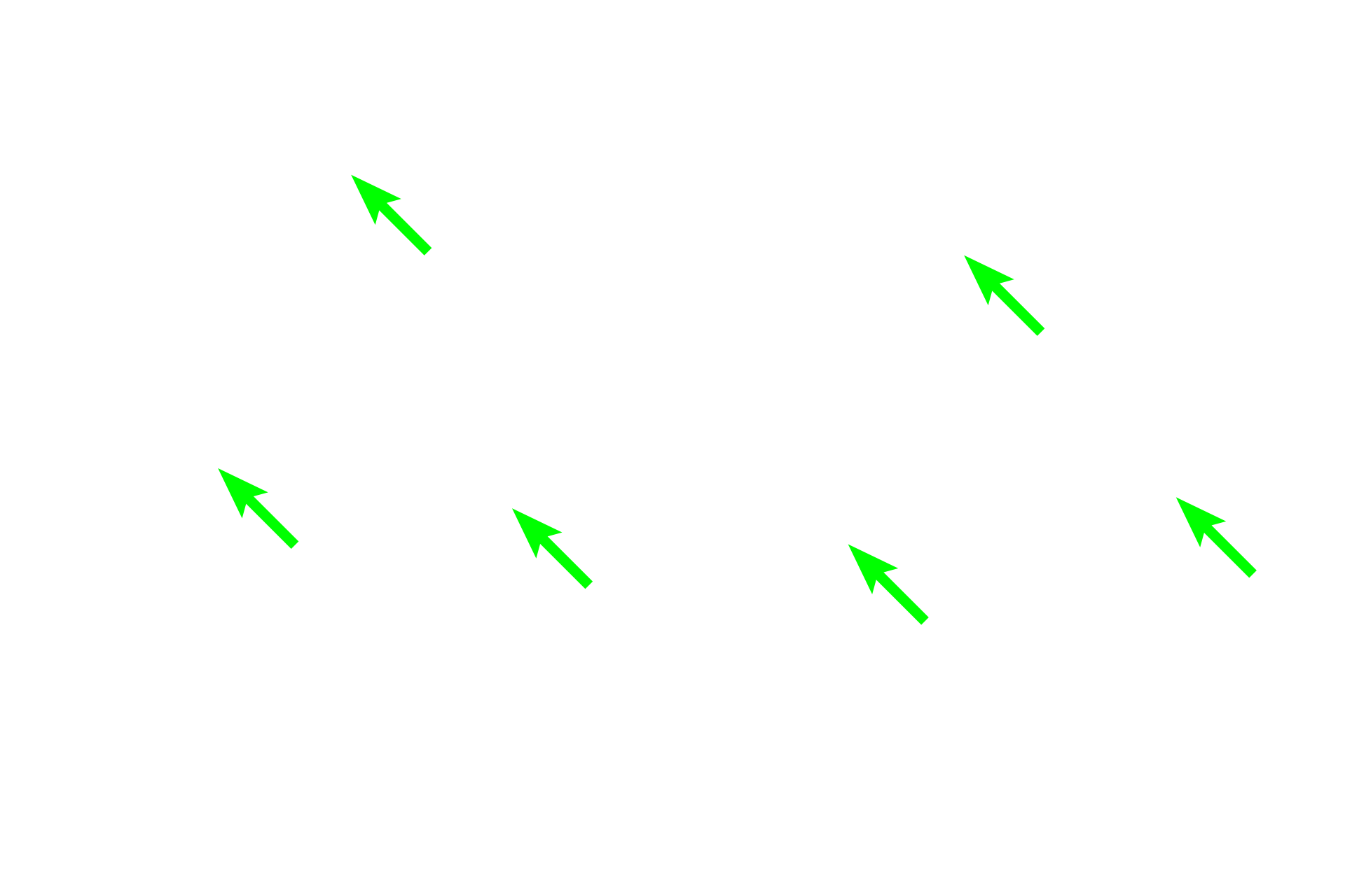
Chrondrocytes
These images compare the appearance of elastic cartilage stained with hematoxylin and eosin (left) and hematoxylin and eosin plus orcein for elastic fibers on the right. 400x
Elastic fibers
These images compare the appearance of elastic cartilage stained with hematoxylin and eosin (left) and hematoxylin and eosin plus orcein for elastic fibers on the right. 400x

Next image
The next image is similar to the area outlined by the rectangle.
Image credit >
This image was taken of a slide in the Drexel University collection.
 PREVIOUS