
Elastic cartilage
Chondrocytes in elastic cartilage are larger and more numerous than in hyaline cartilage and form fewer isogenous groups. The extracellular matrix of elastic cartilage is less abundant than in hyaline cartilage. This special stain demonstrates the abundant elastic fibers in the matrix. Masson stain 100x
Chondrocytes
Chondrocytes in elastic cartilage are larger and more numerous than in hyaline cartilage and form fewer isogenous groups. The extracellular matrix of elastic cartilage is less abundant than in hyaline cartilage. This special stain demonstrates the abundant elastic fibers in the matrix. Masson stain 100x
Isogenous groups
Chondrocytes in elastic cartilage are larger and more numerous than in hyaline cartilage and form fewer isogenous groups. The extracellular matrix of elastic cartilage is less abundant than in hyaline cartilage. This special stain demonstrates the abundant elastic fibers in the matrix. Masson stain 100x
Elastic fibers
Chondrocytes in elastic cartilage are larger and more numerous than in hyaline cartilage and form fewer isogenous groups. The extracellular matrix of elastic cartilage is less abundant than in hyaline cartilage. This special stain demonstrates the abundant elastic fibers in the matrix. Masson stain 100x

Perichondrium >
A perichondrium surrounds elastic cartilage and consists of an outer portion, the fibrous layer, and an inner chondrogenic layer.

- Fibrous layer >
The fibrous layer resembles a dense connective tissue and is a reserve cell layer for the inner layer, the chondrogenic layer. The chondrogenic layer blends imperceptibly with the cartilage, so that its innermost boundary cannot be easily established. Chondroblasts lie in the inner layer immediately adjacent to the cartilage and secrete cartilage matrix around themselves.
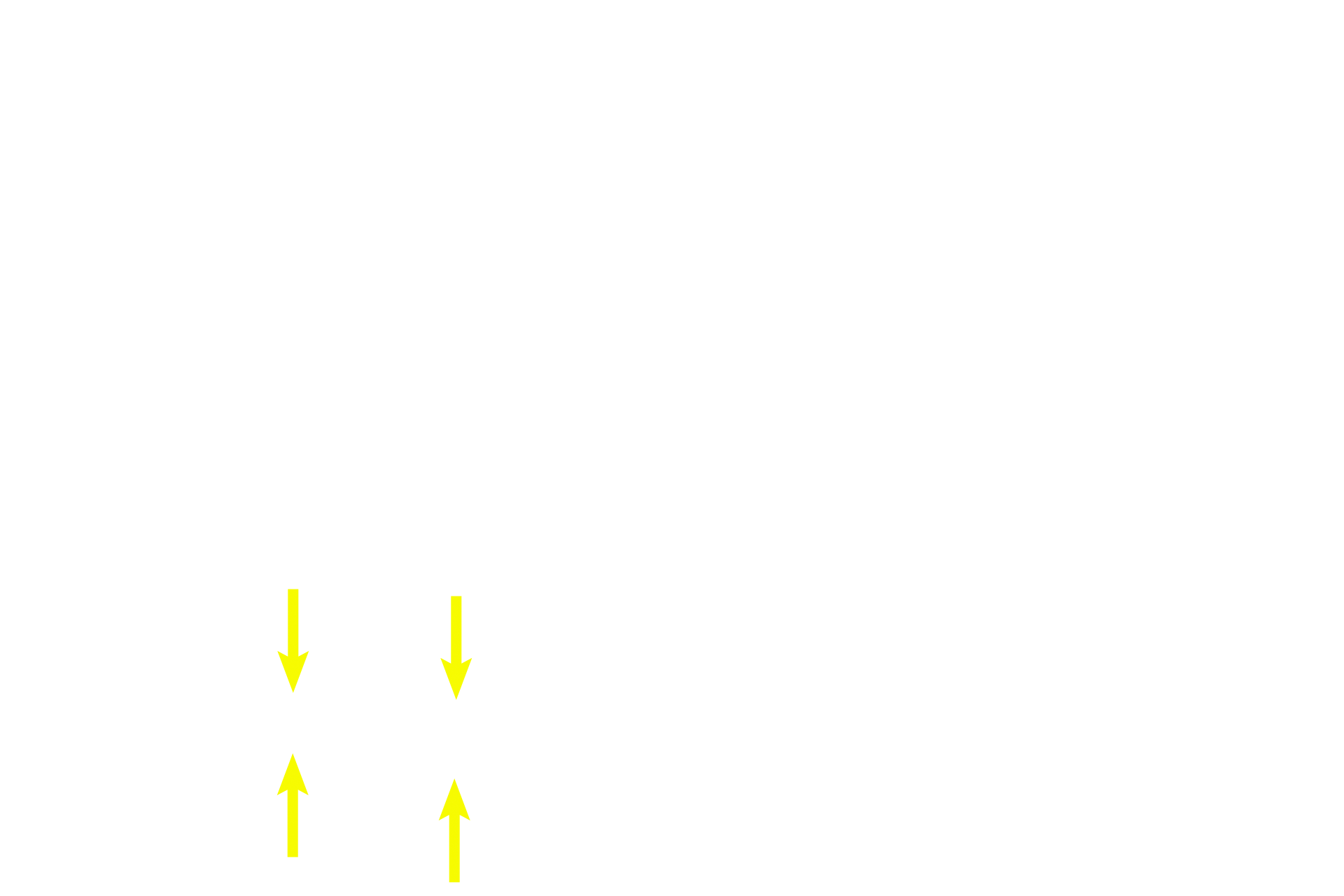
- Chondrogenic layer
The fibrous layer resembles a dense connective tissue and is a reserve cell layer for the inner layer, the chondrogenic layer. The chondrogenic layer blends imperceptibly with the cartilage, so that its innermost boundary cannot be easily established. Chondroblasts lie in the inner layer immediately adjacent to the cartilage and secrete cartilage matrix around themselves.
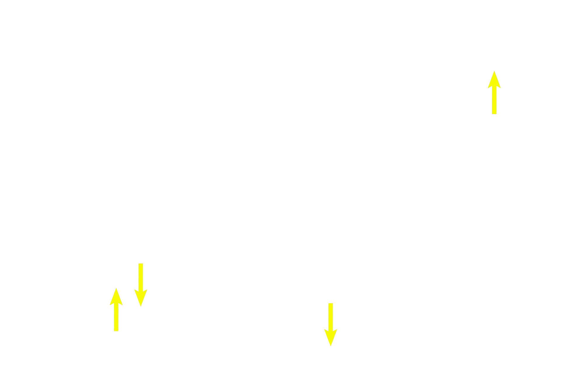
Chondroblasts
A perichondrium surrounds elastic cartilage. The outer portion,the fibrous layer, is a reserve cell layer for the inner layer, the chondrogenic layer. This layer blends imperceptibly with the cartilage, so that its innermost boundary cannot be easily established. Chondroblasts lie in the inner layer immediately adjacent to the cartilage and secrete cartilage matrix around themselves.
Image credit >
This image is taken from a slide in The University of Michigan collection.