
Bone: intramembranous formation
Both images of the fetal skull show the development of spongy woven bone by intramembranous ossification from mesenchyme. Multiple, individual spicules are created at a primary center of ossification. These spicules frequently anastomose and increase in size, forming spongy woven bone. Each spicule is surrounded by a single layer of cells. 400x, 400x
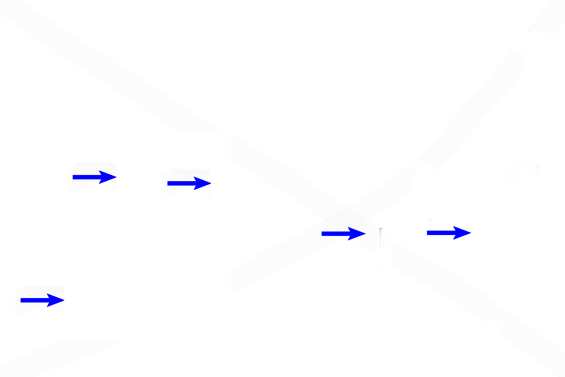
Osteoblasts
Both images of the fetal skull show the development of spongy woven bone by intramembranous ossification from mesenchyme. Multiple, individual spicules are created at a primary center of ossification. These spicules frequently anastomose and increase in size, forming spongy woven bone. Each spicule is surrounded by an endosteum. 400x, 400x
Osteoid
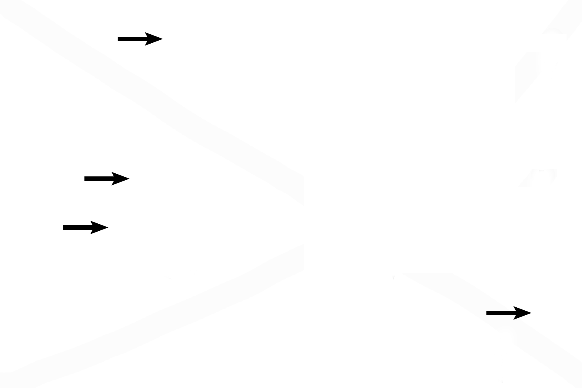
Both images of the fetal skull show the development of spongy woven bone by intramembranous ossification from mesenchyme. Multiple, individual spicules are created at a primary center of ossification. These spicules frequently anastomose and increase in size, forming spongy woven bone. Each spicule is surrounded by an endosteum. 400x, 400x
Lining (endosteal) cells
Both images of the fetal skull show the development of spongy woven bone by intramembranous ossification from mesenchyme. Multiple, individual spicules are created at a primary center of ossification. These spicules frequently anastomose and increase in size, forming spongy woven bone. Each spicule is surrounded by an endosteum. 400x, 400x

Osteocytes
Both images of the fetal skull show the development of spongy woven bone by intramembranous ossification from mesenchyme. Multiple, individual spicules are created at a primary center of ossification. These spicules frequently anastomose and increase in size, forming spongy woven bone. Each spicule is surrounded by an endosteum. 400x, 400x

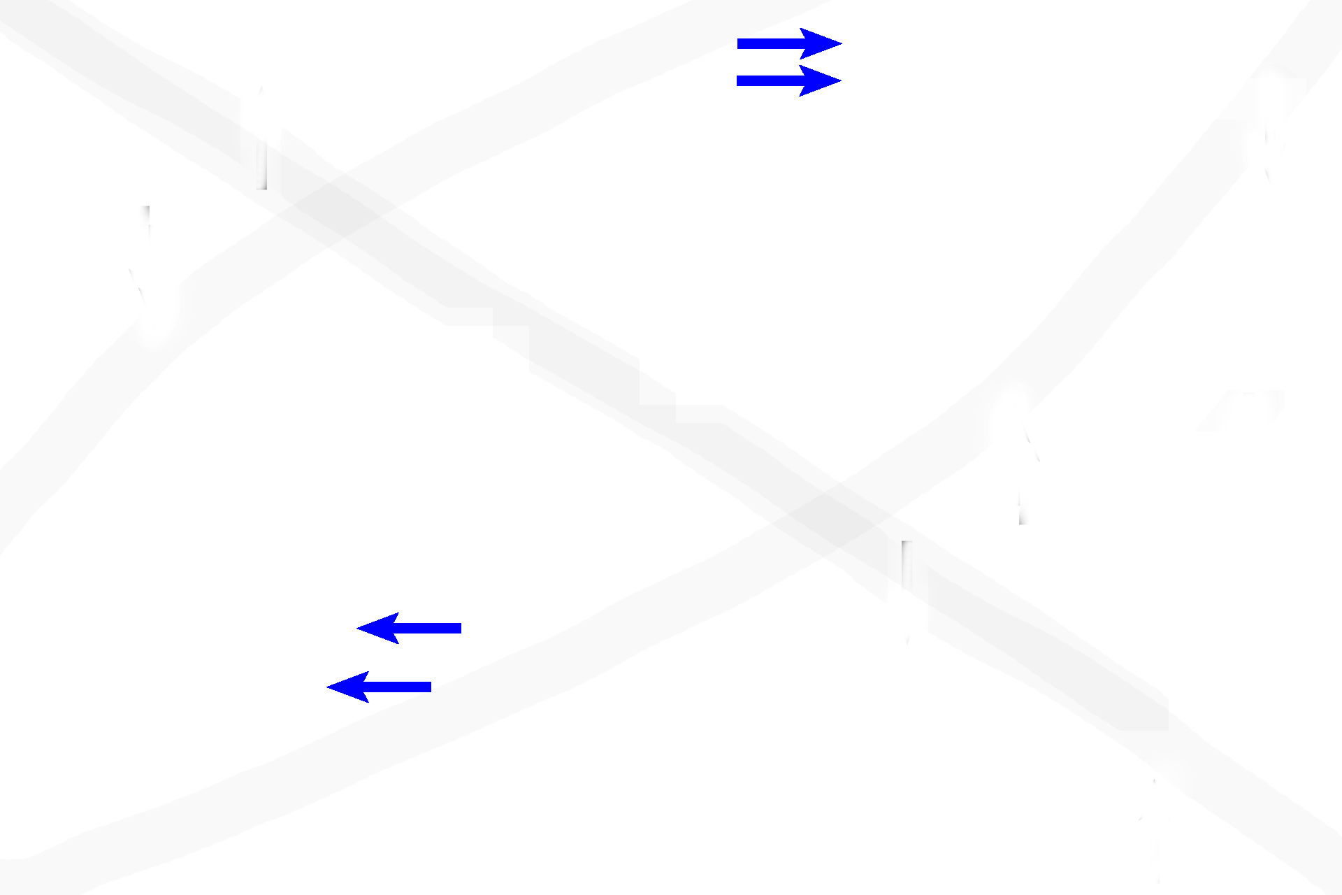
Continued bone formation >
Once initiated by mesenchyme, bone formation continues by the endosteum surrounding the spicules, increasing spicule size. Spicules may merge with each other, as seen in the right image. Imagine how a circular profile (arrow) could be converted to an osteon, thus transforming spongy bone into the compact bone seen in the inner and outer tables of an adult flat bone.

Blood vessels >
Numerous blood vessels are always present within and adjacent to bone.