
Endochondral ossification
A longitudinal section of finger shows endochondral ossification after the primary center of ossification has formed, but before secondary centers in the epiphyses have begun. A periosteal collar surrounds the original primary center. The hyaline cartilage originally located in this center was replaced with bone, which was then resorbed, leaving a marrow cavity in its place. 40x

Hyaline cartilage >
Typical hyaline cartilage formed the original bony template and still exists in the region of the future epiphyses.
Periosteal collar >
Early in endochondral ossification, the perichondrium becomes vascularized, enabling this layer to act as a periosteum and deposit bone at the periphery of the cartilage template. This periosteal collar is formed by intramembranous ossification, as bone is deposited on, rather than replacing, cartilage. The periosteal collar supports forming bone and contributes to its future diaphysis.
Marrow cavity >
At the primary center of ossification, chondrocytes matured, hypertrophied and regressed, producing calcified cartilage spicules in the process. Bone was deposited on the spicules, but both cartilage and bone were later resorbed, producing a marrow space.

Epiphyseal plates >
Chondrocytes at both ends of the marrow cavity align in rows forming epiphyseal plates, which continue the regressive changes. The bone deposition and resorption that follow, add length to the developing bone.
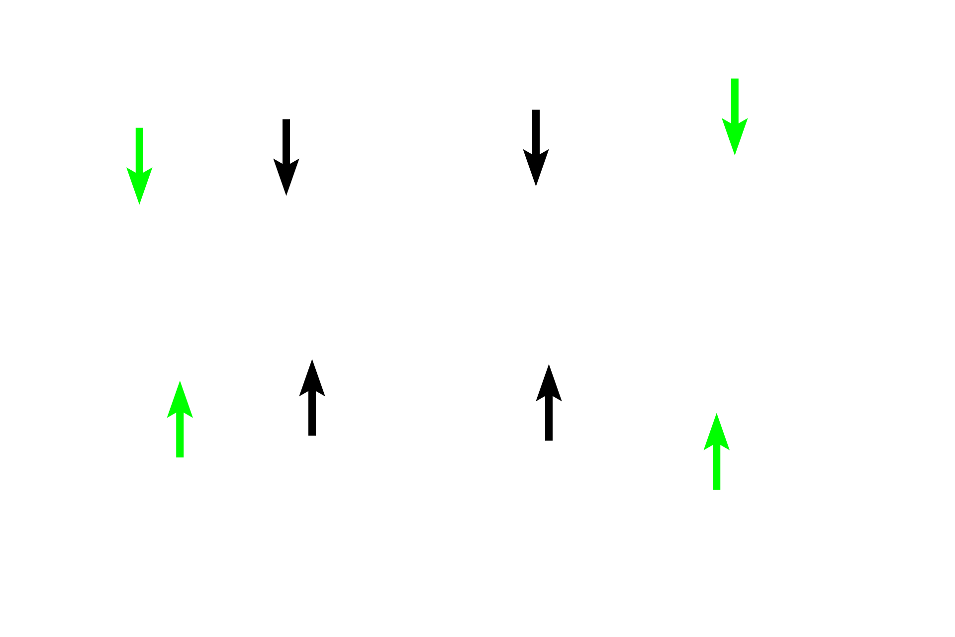
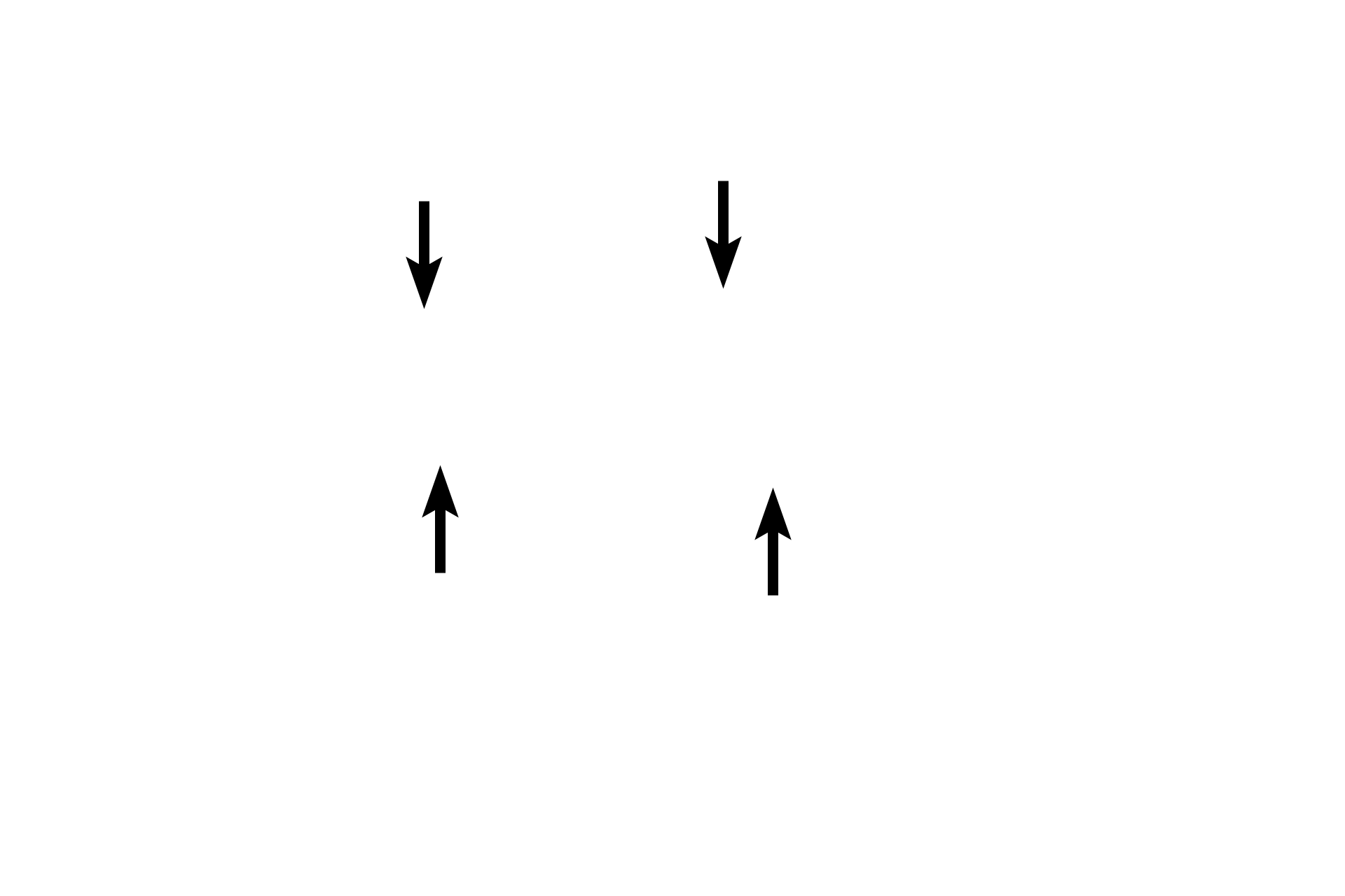
Perichondrium/periosteum >
The original, fetal perichondrium (green arrows) continues to cover the hyaline cartilage, while the continuation of the perichondrium over the periosteal band is now the periosteum (black arrows). The periosteum is responsible for the production of the periosteal band.

Next image >
The next image is similar to that seen in the rectangle.