
Endochondral ossification
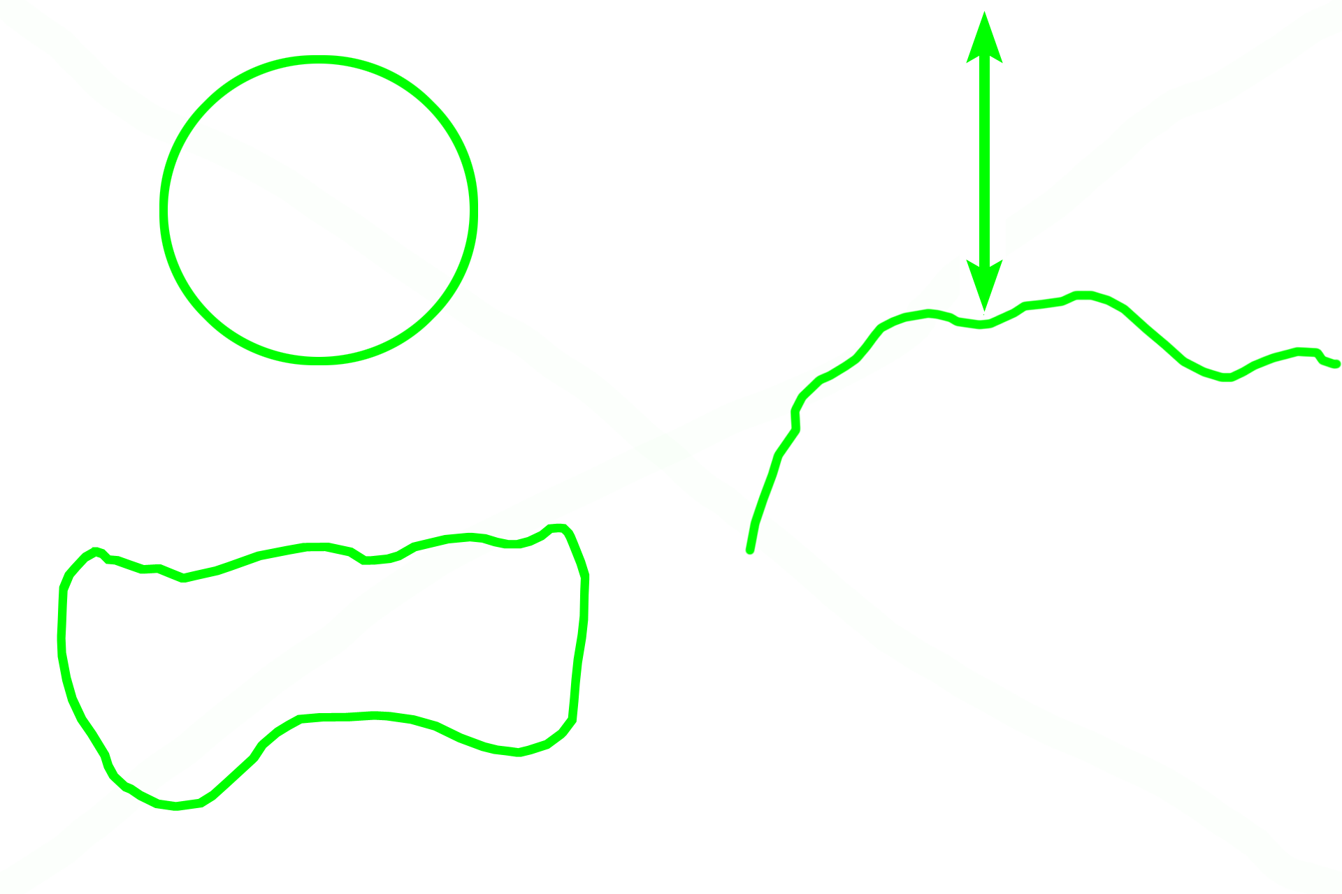
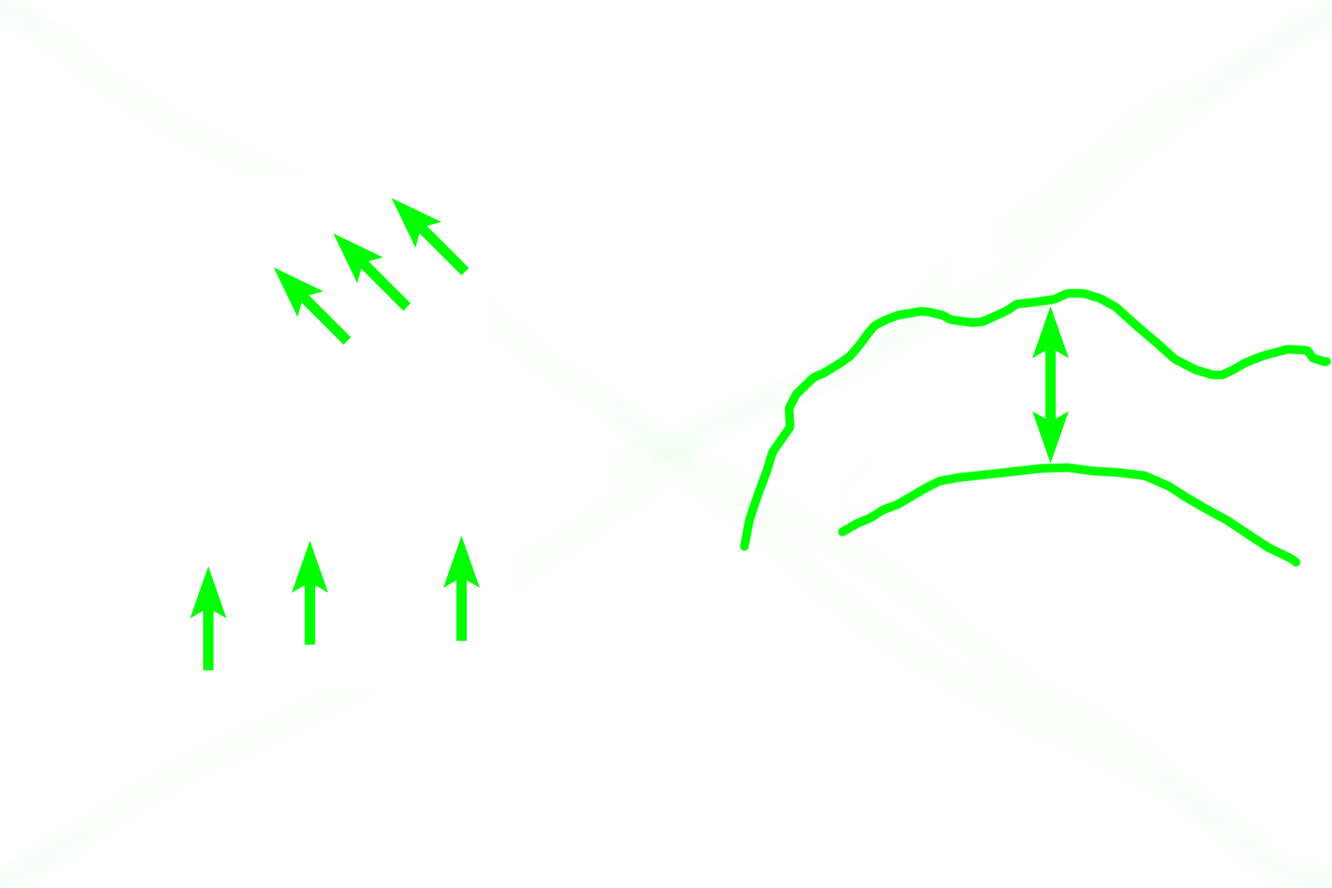
On the left are images of the proximal (upper) and distal (lower) ends of the femur, showing two epiphyseal plates in this long bone. On the right, a decalcified section of another long bone, the humerus, demonstrates its epiphyseal plate at low magnification. 10x
Epiphysis >
The epiphysis is the terminal enlargement of a long bone. Its periphery is formed of a thin layer of compact bone, while the interior is spongy bone and red marrow. The proximal epiphysis of the femur has been modified to form a head and neck.
Epiphyseal plate >
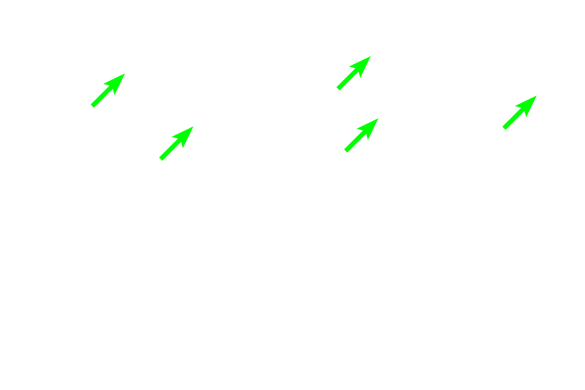
Growth in length occurs from a region of hyaline cartilage, the epiphyseal plate. The locations of two epiphyseal plates (green arrows) are still present in the left images. After chondrocyte proliferation stops, further growth in length ceases and only an epiphyseal line remains to indicate the original location of the plate.
Metaphysis >
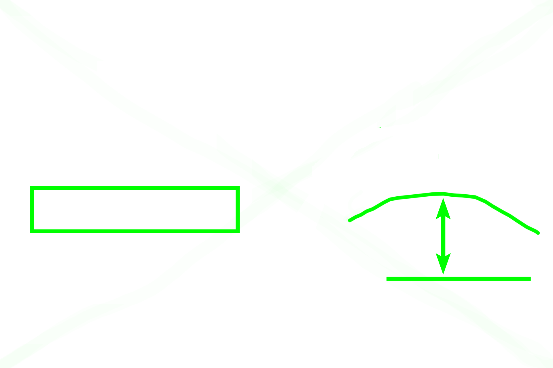
The metaphysis is the funnel-shaped segment of bone interposed between the diaphysis and the epiphyseal plate (if the bone is still growing in length) or the epiphyseal line and adjacent epiphysis (if growth has ceased).

Diaphysis >
The diaphysis of the bone is composed of compact bone and is filled with yellow marrow in the adult.
Compact bone >
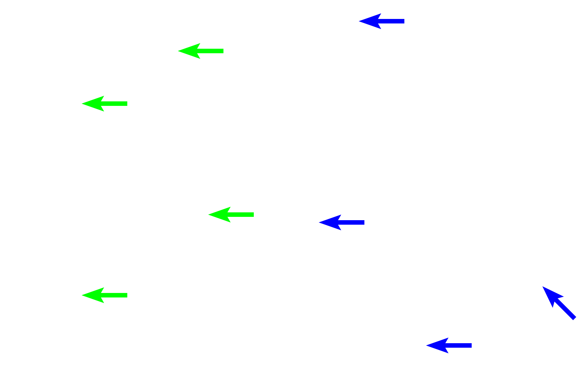
Compact bone covers the periphery of the entire bone and forms the thick bone comprising the diaphysis of a long bone.
Spongy bone >
Spongy bone is found in the interior of the bone, filling the epiphyses and lining the compact bone in the diaphysis. Red marrow fills the spaces of spongy bone in the epiphyses.

Red marrow
Spongy bone is found in the interior of the bone, filling the epiphyses and lining the compact bone in the diaphysis. Red marrow fills the spaces of spongy bone in the epiphyses.
 PREVIOUS