
Bone: the organ - articular cartilage
The ends of long bones are covered by articular cartilage, formed of hyaline cartilage, providing a smooth, glassy surface that allows the ends of the bones to move easily on each other. This type of articulation is called a synovial joint. Articular cartilages, which are not covered by periosteum, are separated by a fluid-filled synovial space. A thin layer of compact bone lies beneath the cartilage. 10x, 800x

Epiphyses
The ends of long bones are covered by articular cartilage, formed of hyaline cartilage, providing a smooth, glassy surface that allows the ends of the bones to move easily on each other. This type of articulation is called a synovial joint. Articular cartilages, which are not covered by periosteum, are separated by a fluid-filled synovial space. A thin layer of compact bone lies beneath the cartilage. 10x, 800x

Articular cartilages
The ends of long bones are covered by articular cartilage, formed of hyaline cartilage, providing a smooth, glassy surface that allows the ends of the bones to move easily on each other. This type of articulation is called a synovial joint. Articular cartilages, which are not covered by periosteum, are separated by a fluid-filled synovial space. A thin layer of compact bone lies beneath the cartilage. 10x, 800x

- Chondrocytes
The ends of long bones are covered by articular cartilage, formed of hyaline cartilage, providing a smooth, glassy surface that allows the ends of the bones to move easily on each other. This type of articulation is called a synovial joint. Articular cartilages, which are not covered by periosteum, are separated by a fluid-filled synovial space. A thin layer of compact bone lies beneath the cartilage. 10x, 800x
Compact bone
The ends of long bones are covered by articular cartilage, formed of hyaline cartilage, providing a smooth, glassy surface that allows the ends of the bones to move easily on each other. This type of articulation is called a synovial joint. Articular cartilages, which are not covered by periosteum, are separated by a fluid-filled synovial space. A thin layer of compact bone lies beneath the cartilage. 10x, 800x
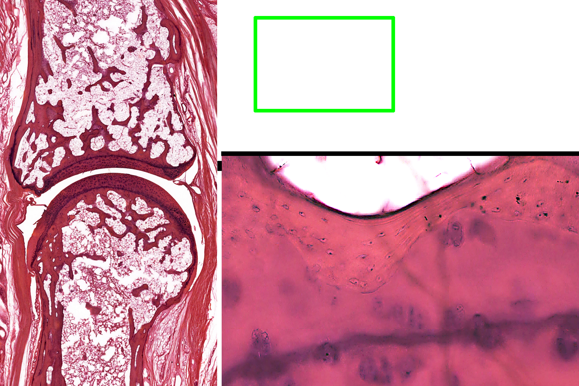
Bone-cartilage interface >
This higher magnification of the area indicated by the green rectangle shows the interface of the compact bone of the epiphysis with articular cartilage. Osteocytes in lacuna are evident in the compact bone.
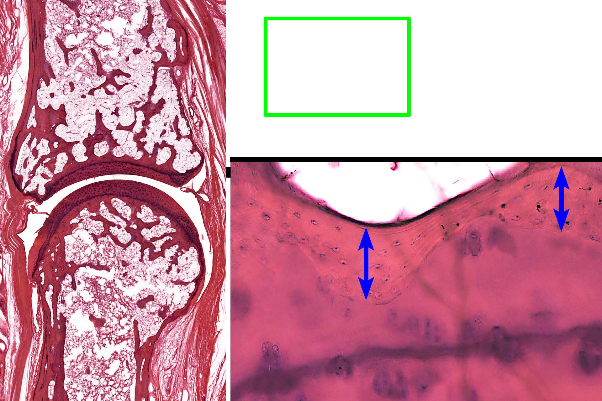
- Bone
This higher magnification of the area indicated by the green rectangle shows the interface of the compact bone of the epiphysis with articular cartilage. Osteocytes in lacuna are evident in the compact bone.
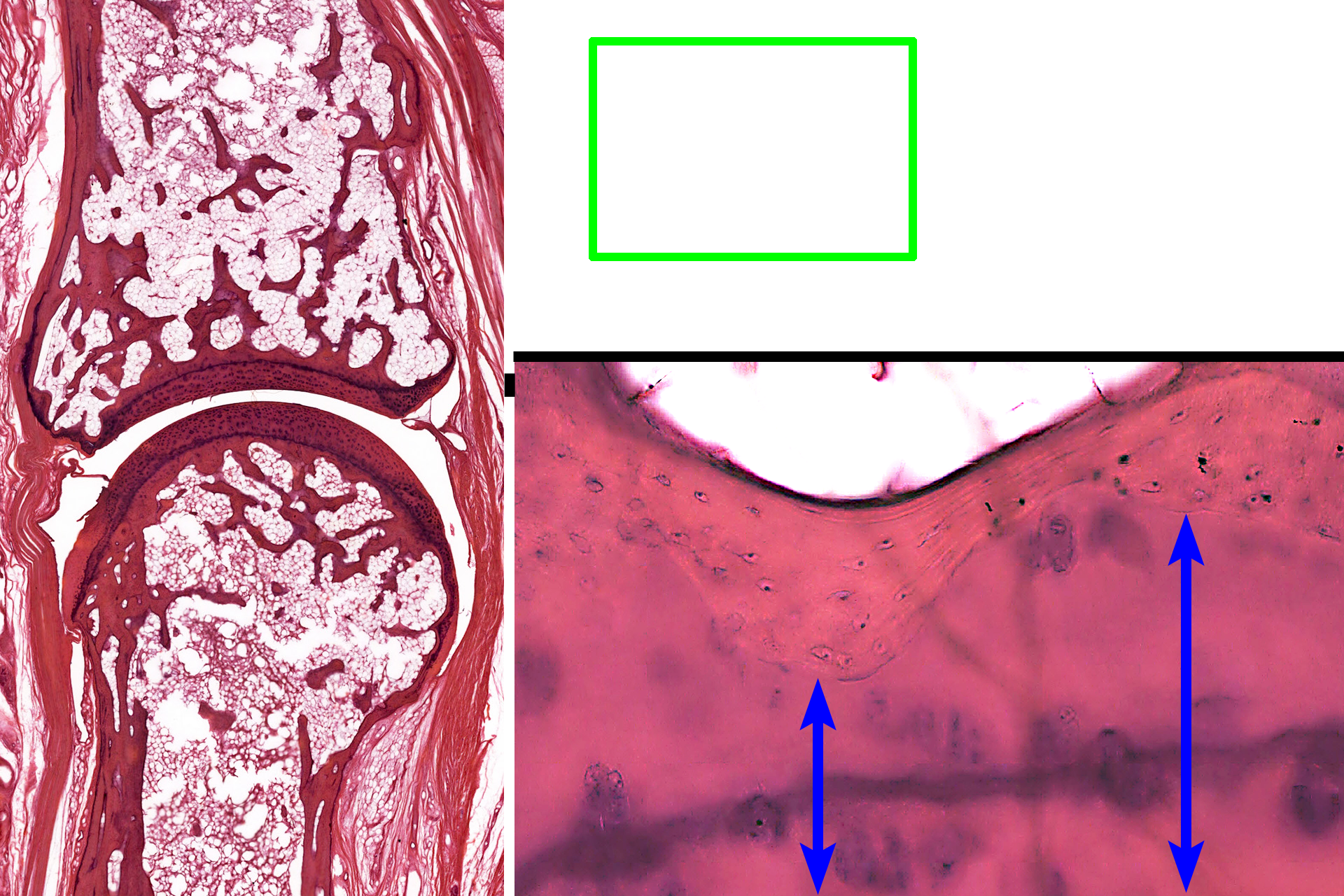
- Cartilage
This higher magnification of the area indicated by the green rectangle shows the interface of the compact bone of the epiphysis with articular cartilage. Osteocytes in lacuna are evident in the compact bone.

Spongy bone
The ends of long bones are covered by articular cartilage, formed of hyaline cartilage, providing a smooth, glassy surface that allows the ends of the bones to move easily on each other. This type of articulation is called a synovial joint. Articular cartilages, which are not covered by periosteum, are separated by a fluid-filled synovial space. A thin layer of compact bone lies beneath the cartilage. 10x, 800x

Synovial space
The ends of long bones are covered by articular cartilage, formed of hyaline cartilage, providing a smooth, glassy surface that allows the ends of the bones to move easily on each other. This type of articulation is called a synovial joint. Articular cartilages, which are not covered by periosteum, are separated by a fluid-filled synovial space. A thin layer of compact bone lies beneath the cartilage. 10x, 800x
Image source >
Images taken of a slide in the University of New England College of Osteopathic Medicine slide collection.