
Bone: macroscopic appearance
The femur above and the drawing of the humerus below illustrate bone as an organ. The organ bone is composed of bone tissue, its internal and external coverings, the blood and nerve supplies, marrow, and articular cartilage(s). The subdivisions of a long bone are illustrated here also.
Subdivisions >
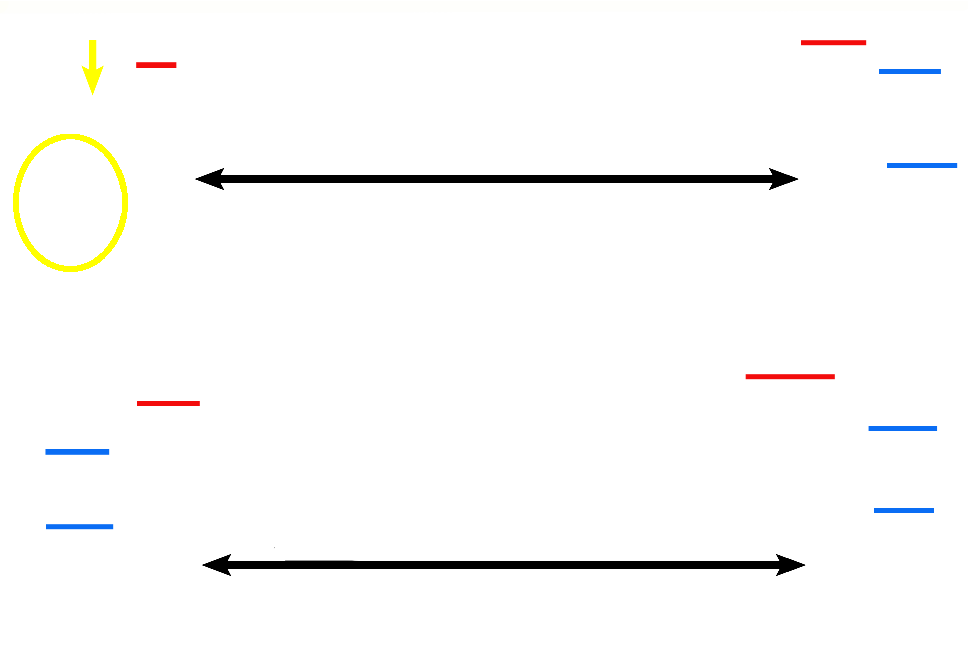
The shaft (black arrows) of a long bone is called the diaphysis. The “knob” at either end of the bone is an epiphysis (blue bars). One epiphysis (left) of the femur is modified to form the greater trochanter (yellow arrow) and the head and neck (yellow circle). The wedge-shaped subdivision separating the diaphysis from each epiphysis is the metaphysis (red bars).

Periosteum >
Because this femur was not fixed for histological viewing and because it was not included in the drawing, no living tissue is demonstrable here. Therefore, the outer covering of bone, the periosteum, whose position is indicated by the arrows, is absent. The periosteum covers all external surfaces of bone except where articular cartilages are present.

Endosteum >
The endosteum, like the periosteum, was not preserved in this preparation and must be imagined. The endosteum is a single layer of cells that lines all interior bony surfaces except lacunae and canaliculi.
Marrow >
Red marrow (red arrow and red bars) fills spongy bone areas of bones; red marrow is myeloid tissue that produces red and white blood cells. Yellow marrow (blue arrows) is composed of adipose connective tissue and myeloid tissue. Yellow marrow has the ability to convert to red marrow, if needed, and is located in the center of the diaphysis of a long bone.

Articular cartilages >
Articular cartilage is hyaline cartilage that covers the articulating surfaces of bone. The smooth, glassy surface of this cartilage allows the ends of the bones to move easily on each other. No periosteum is present on articular cartilage.
Epiphyseal plates >
Long bone grows in length from a plate of hyaline cartilage, called an epiphyseal plate, located between the epiphysis and the metaphysis. Growth had not stopped in this femur so epiphyseal plates remain. Because the organic, extracellular bony components were not preserved in this femur, epiphyseal plates are represented only by the narrow gaps where the plates (red arrows) were located.
Epiphyseal line >
Growth stops when the epiphyseal plate closes, i.e., stops functioning, leaving an epiphyseal line in its place.
Compact bone >
Compact bone covers all exterior surfaces of bone and forms the majority of the thick wall of the diaphysis. Spongy bone is located in the interior of bones and can be seen in the epiphyseal and metaphyseal regions. Spongy bone also forms a thin lining around the interior of the compact bone of the shaft.

Spongy bone
Compact bone covers all exterior surfaces of bone and forms the majority of the thick wall of the diaphysis. Spongy bone is located in the interior of bones and can be seen in the epiphyseal and metaphyseal regions. Spongy bone also forms a thin lining around the interior of the compact bone of the shaft.