
Renal calyx
These images show the structure of the renal calyx (left) in a uni-pyramidal kidney, and the mucosa of the calyx at higher magnification (right). A calyx surrounds each pyramid, and its mucosa is reflected onto the conical surface of the pyramid. 100x, 800x.
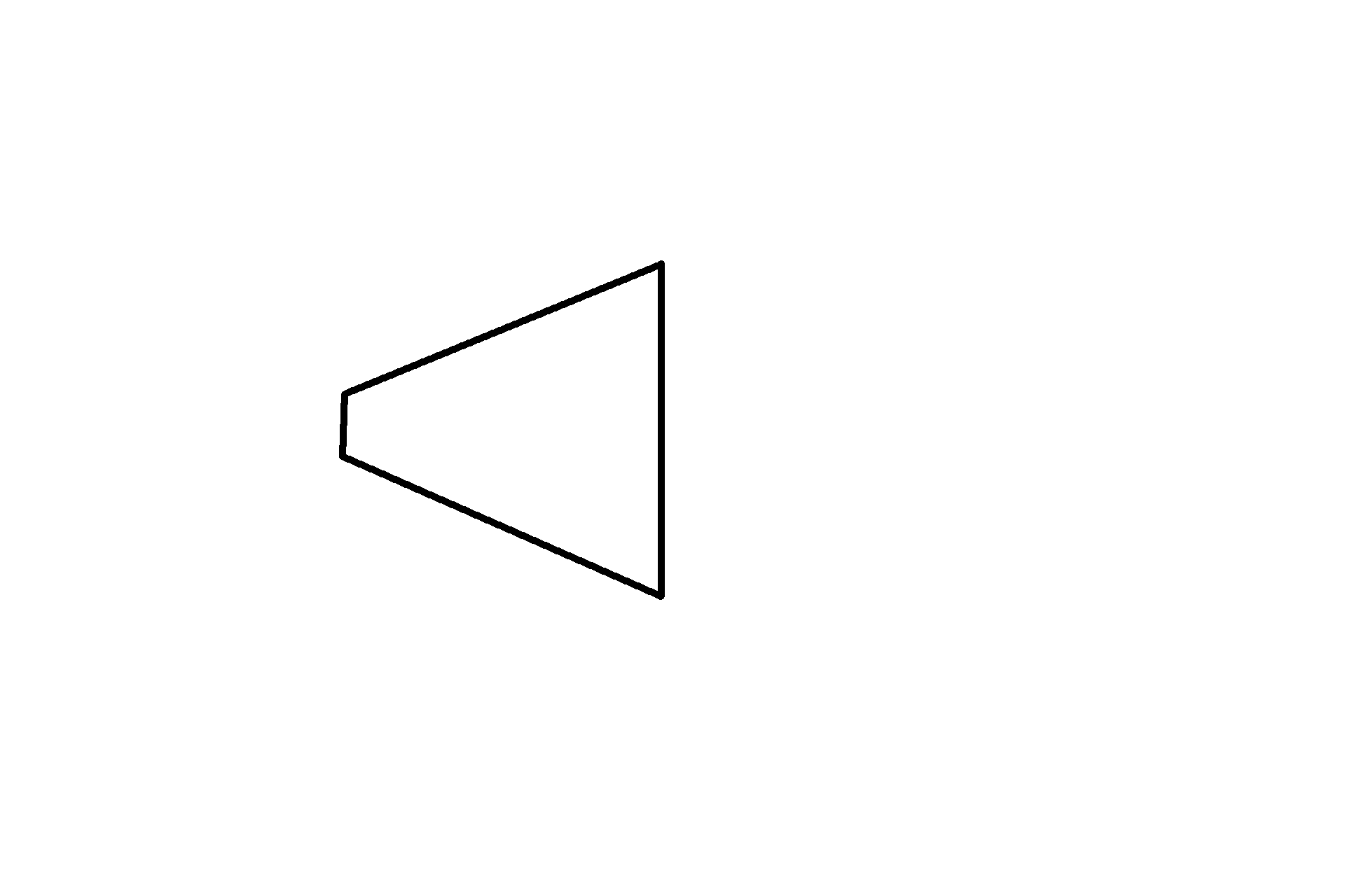
Renal papilla >
The renal pyramid is a conical-shaped structure, but only its apex, the papilla (outlined), is visible in this image. The papilla of the pyramid receives the terminal ends of the papillary ducts of Bellini. The number of renal pyramids varies between species: humans have 8-10 pyramids. This image shows an example of a uni-pyramidal kidney.

- Area cribosa >
The pointed end of the renal papilla is referred to as the area cribosa. Here, the terminations of the papillary ducts of Belllini empty into the lumen of the calyx.

Calyx >
The calyx, a chalice-like covering over the papilla, receives urine from the papillary ducts of Bellini. The transitional epithelium of the calyx (arrows) is reflected onto the surface of the papilla to enclose the lumen of the calyx (filled in blue).

- Calyx mucosa >
The mucosa of the calyx consists of a transitional epithelium with its underlying, thin lamina propria and two layers of smooth muscle oriented perpendicularly to each other.
- Transitional epithelium
The mucosa of the calyx consists of a transitional epithelium with its underlying, thin lamina propria and two layers of smooth muscle oriented perpendicularly to each other.
- Lamina propria
The mucosa of the calyx consists of a transitional epithelium with its underlying, thin lamina propria and two layers of smooth muscle oriented perpendicularly to each other.
- Smooth muscle
The mucosa of the calyx consists of a transitional epithelium with its underlying, thin lamina propria and two layers of smooth muscle oriented perpendicularly to each other.

Renal sinus >
The renal sinus is the space at the indented hilum of the kidney. The excretory passageways and major blood vessels are located within the sinus. The sinus is filled with adipose and loose connective tissues.

- Renal pelvis >
The calyces merge to eventually form the renal pelvis, a portion of which is visible here. The renal pelvis is the expanded proximal portion of the ureter and lies in the renal sinus along with the major renal blood vessels.

- Renal blood vessels
The calyces merge to eventually form the renal pelvis, a portion of which is visible here. The renal pelvis is the expanded proximal portion of the ureter and lies in the renal sinus along with the major renal blood vessels.

Image source >
This image was taken from a slide in the Drexel University slide collection.