Renal pyramid
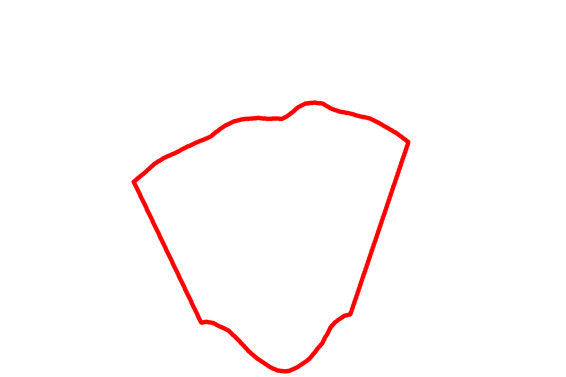
This section of non-primate kidney shows a renal pyramid with its papilla inserted into a minor calyx. Human kidneys have 10-18 pyramids, in contrast to this example of a uni-pyramidal kidney. Collecting ducts descend from the medullary rays, traverse the medulla and converge at the apex of the papilla as papillary ducts of Bellini, where they deliver urine into the lumen of the minor calyx. 10x

Capsule >
The kidney is covered by a connective tissue capsule which passes inward at the hilum and becomes continuous with the connective tissue forming the walls of the calyces.

Cortex >
The cortex consists of convoluted regions (renal corpuscles, convoluted regions of proximal and distal tubules, and connecting tubules) and medullary rays (straight portions of proximal and distal tubules and cortical collecting ducts). The cortex is divided into cortical renal lobules, each of which has a medullary ray at its center and interlobular arteries in the convoluted portion at its perimeter.

- Convoluted portions
The cortex consists of convoluted regions (renal corpuscles, convoluted regions of proximal and distal tubules, and connecting tubules) and medullary rays (straight portions of proximal and distal tubules and cortical collecting ducts). The cortex is divided into cortical renal lobules, each of which has a medullary ray at its center and interlobular arteries in the convoluted portion at its perimeter.

- Medullary rays
The cortex consists of convoluted regions (renal corpuscles, convoluted regions of proximal and distal tubules, and connecting tubules) and medullary rays (straight portions of proximal and distal tubules and cortical collecting ducts). The cortex is divided into cortical renal lobules, each of which has a medullary ray at its center and interlobular arteries in the convoluted portion at its perimeter.

- Renal lobules
The cortex consists of convoluted regions (renal corpuscles, convoluted regions of proximal and distal tubules, and connecting tubules) and medullary rays (straight portions of proximal and distal tubules and cortical collecting ducts). The cortex is divided into cortical renal lobules, each of which has a medullary ray at its center and interlobular arteries in the convoluted portion at its perimeter.

Medulla >
The medulla is subdivided into outer and inner regions, with the outer medulla being further subdivided into an inner stripe and an outer stripe. The variation in staining visible in these regions reflects the location of distinct parts of the uriniferous tubule at different levels of the pyramid.

- Outer medulla
The medulla is subdivided into outer and inner regions, with the outer medulla being further subdivided into an inner stripe and an outer stripe. The variation in staining visible in these regions reflects the location of distinct parts of the uriniferous tubule at different levels of the pyramid.

-- Outer stripe
The medulla is subdivided into outer and inner regions, with the outer medulla being further subdivided into an inner stripe and an outer stripe. The variation in staining visible in these regions reflects the location of distinct parts of the uriniferous tubule at different levels of the pyramid.

-- Inner stripe
The medulla is subdivided into outer and inner regions, with the outer medulla being further subdivided into an inner stripe and an outer stripe. The variation in staining visible in these regions reflects the location of distinct parts of the uriniferous tubule at different levels of the pyramid.

- Inner medulla
The medulla is subdivided into outer and inner regions, with the outer medulla being further subdivided into an inner stripe and an outer stripe. The variation in staining visible in these regions reflects the location of distinct parts of the uriniferous tubule at different levels of the pyramid.

Pyramid >
Renal pyramids are conical structures composed of the straight, parallel segments of nephrons and collecting ducts. The base of a pyramid lies adjacent to the cortex, while its apex (papilla) projects into a minor calyx. A renal lobe is defined as a renal pyramid and its adjacent and overlying cortical tissue. Human kidneys have 10-18 lobes, in contrast to this example of a unilobar (uni-pyramidal) kidney.

Arcuate vessels >
The location of the arcuate vessels demarcate the cortex from the medulla. Branches of the arcuate artery, interlobular arteries, ascend into the cortex where they give off afferent arterioles to supply the glomerulus.

Papillary ducts >
Papillary ducts of Bellini are the last portions of the uriniferous tubule and open at the papilla of the pyramid, a region called the area cribosa.

Minor calyx >
Urine draining from the papillary ducts flows into the minor calyx.

Area shown in next image
This area is shown at higher magnification in the next image

Image source >
Image taken from a slide in the University of New England College of Osteopathic Medicine collection.