
Skin overview
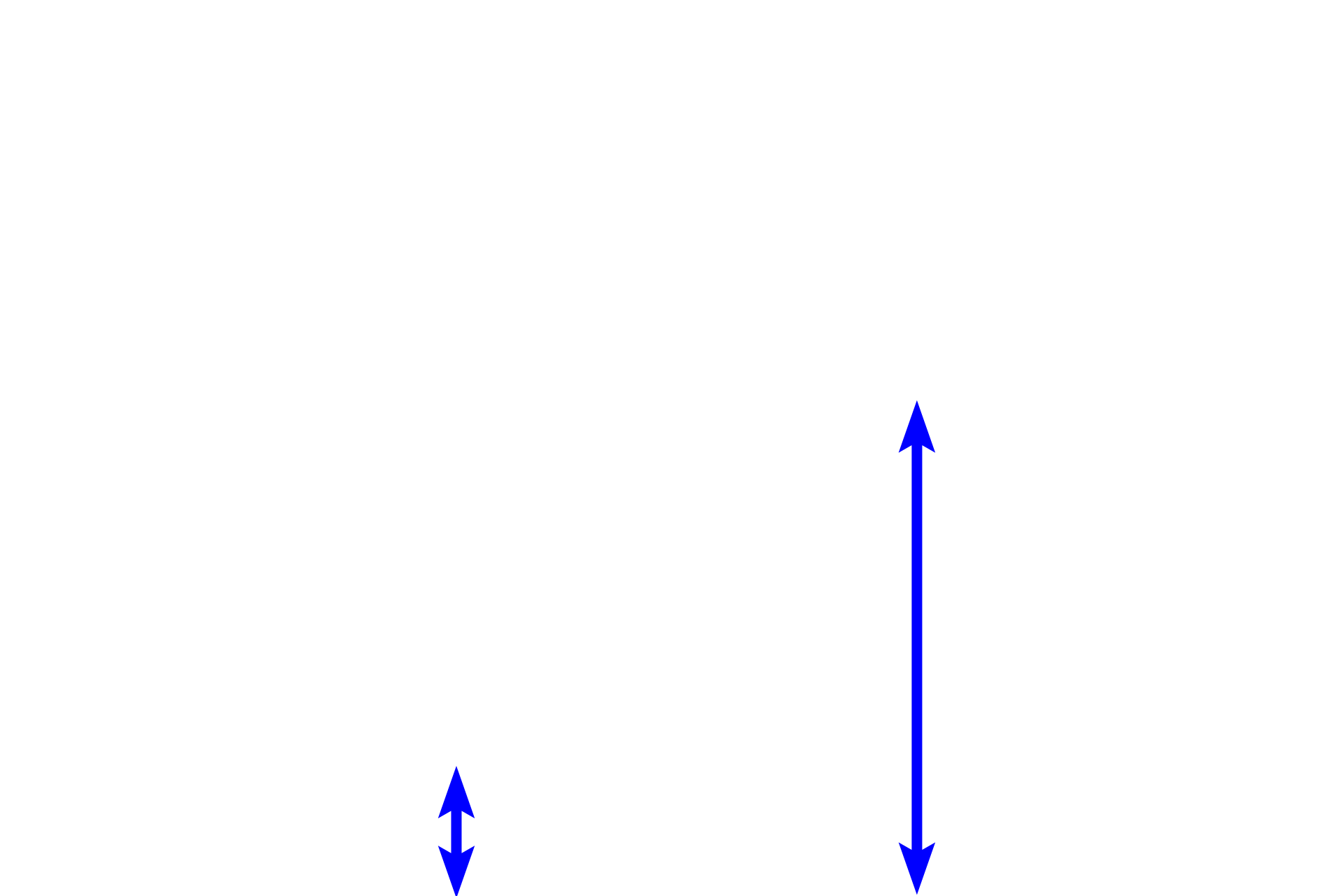
Illustrations of thick skin on the left and thin skin on the right demonstrate the organization of keratinocytes into five layers, or strata. Beginning with the deepest layer, the strata are: basale (germinativum), spinosum, granulosum, lucidum and corneum. Most of these layers are thinner in thin skin and, in some cases, may not be evident as distinct layers.
Stratum basale >
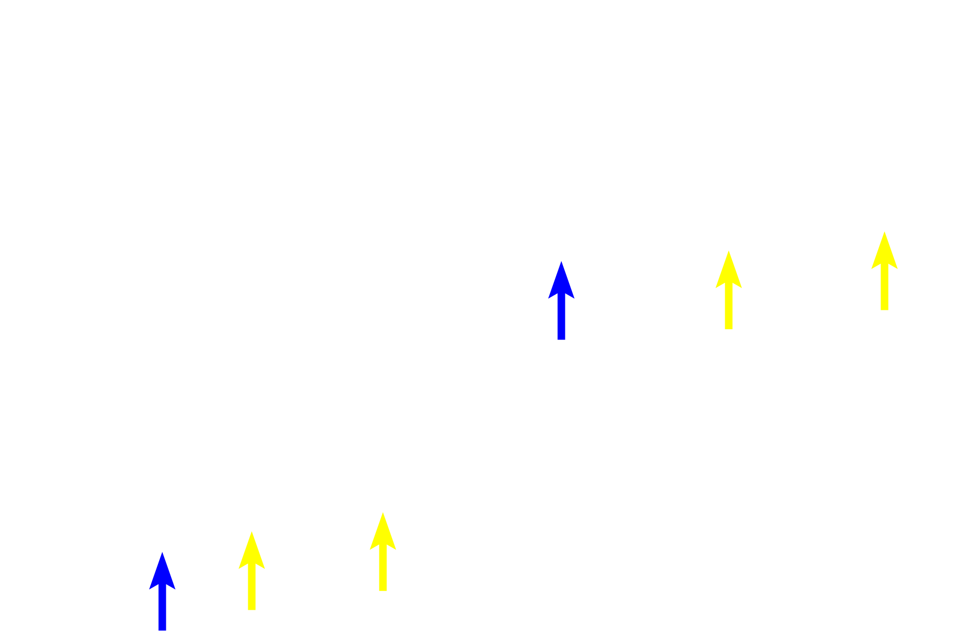
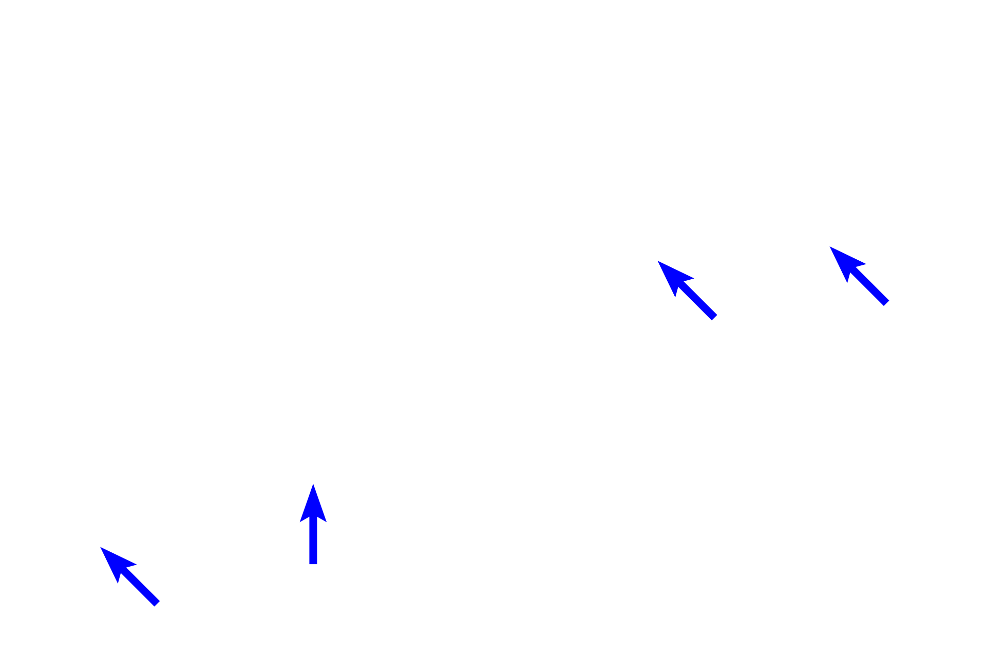
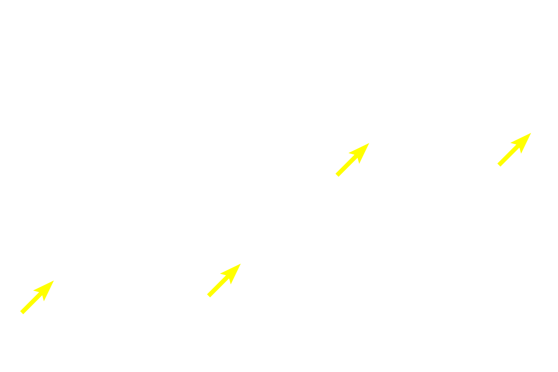
Stratum basale (or germinativum, yellow arrows) consists of two cell types. A single row of cuboidal or columnar keratinocytes rest on the basement membrane separating epidermis from dermis. As its name indicates, intense mitotic activity occurs in this layer to replenish the epidermis. A second cell type, melanocytes (blue arrows), produce melanin and appear as pale cells within stratum basale.
Stratum spinosum >
Stratum spinosum possesses 3-10 layers of keratinocytes, the deepest of which also show mitotic activity. Cells in this layer are joined by numerous desmosomes, creating the appearance of spiny processes around their entire peripheries. The number of layers of stratum spinosum is greatly reduced in thin skin compared to thick skin.
Stratum granulosum >
Stratum granulosum consists of three to five cell layers in thick skin and only one or two layers in thin skin. The prominent, basophilic granules present in these keratinocytes contribute to the formation of keratin.

Stratum lucidum >
Stratum lucidum is a thin translucent layer that is visible only in thick skin on the palms and soles of the feet. It is not present as a distinct layer elsewhere and is often considered to be a subdivision of the stratum corneum. The stratum lucidum lacks cell nuclei and organelles.
Stratum corneum >
Stratum corneum represents the flattened squames of keratinocytes that are entirely filled with the scleroprotein, keratin. Stratum corneum is quite thick in thick skin but is greatly reduced in thin skin.

Dermis >
The dermis, lying beneath the epidermis, consists of two layers. Immediately beneath the epidermis is the papillary layer, composed of loose connective tissue. Beneath that is the reticular layer, composed of dense irregular connective tissue. These two layers blend at their junction, with no distinct demarcation between the two layers.
- Papillary layer of dermis
The dermis, lying beneath the epidermis, consists of two layers. Immediately beneath the epidermis is the papillary layer, composed of loose connective tissue. Beneath that is the reticular layer, composed of dense irregular connective tissue. These two layers blend at their junction, with no distinct demarcation between the two layers.
- Reticular layer of dermis
The dermis, lying beneath the epidermis, consists of two layers. Immediately beneath the epidermis is the papillary layer, composed of loose connective tissue. Beneath that is the reticular layer, composed of dense irregular connective tissue. These two layers blend at their junction, with no distinct demarcation between the two layers.

Epidermal ridges (pegs) >
The interface between epidermis and dermis is strengthened by an undulating surface of downwardly extending epidermal ridges or pegs (rete ridges or pegs) that interdigitate with upwardly projecting dermal papillae. The depth of these interdigitations is greater in thick than in thin skin, reflecting the need for increased resistance against friction and stress on the palms of the hands and soles of the feet.
Dermal papillae
The interface between epidermis and dermis is strengthened by an undulating surface of downwardly extending epidermal ridges or pegs (rete ridges or pegs) that interdigitate with upwardly projecting dermal papillae. The depth of these interdigitations is greater in thick than in thin skin, reflecting the need for increased resistance against friction and stress on the palms of the hands and soles of the feet.

Keratinization >
Keratinocytes in stratum basale produce intermediate filaments (tonofilaments) composed of the protein keratin. These filaments aggregate to form tonofibrils, prominent in stratum spinosum. In stratum granulosum keratohyalin granules and tonofibrils condense to form keratin; simultaneously, organelles and nuclei are lost. The resultant, flattened, keratin-filled squames form stratum corneum.