
Stratum corneum
This electron micrograph shows the outermost layers of cells of the stratum corneum with two desquamated cells at its surface. Each keratinized cell is less than one micron thick and is filled with an amorphous matrix containing aggregated keratin filaments. Desmosomal junctions are mostly degraded in the upper regions of the stratum corneum. 15,000x
Stratum corneum cells
This electron micrograph shows the outermost layers of cells of the stratum corneum with two desquamated cells at its surface. Each keratinized cell is less than one micron thick and is filled with an amorphous matrix containing aggregated keratin filaments. Desmosomal junctions are mostly degraded in the upper regions of the stratum corneum. 15,000x

Desquamated cells
This electron micrograph shows the outermost layers of cells of the stratum corneum with two desquamated cells at its surface. Each keratinized cell is less than one micron thick and is filled with an amorphous matrix containing aggregated keratin filaments. Desmosomal junctions are mostly degraded in the upper regions of the stratum corneum. 15,000x
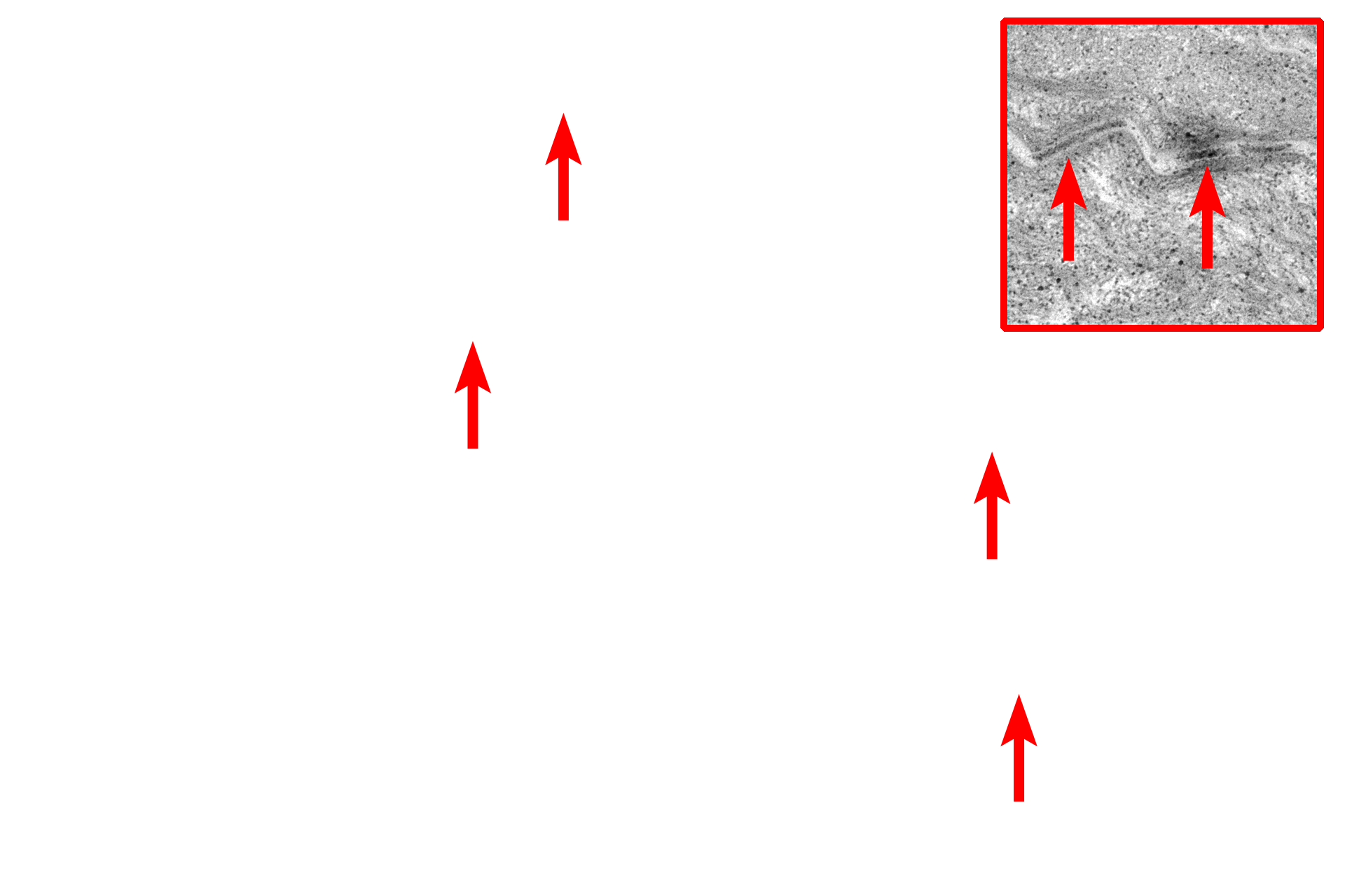
Desmosomes
This electron micrograph shows the outermost layers of cells of the stratum corneum with two desquamated cells at its surface. Each keratinized cell is less than one micron thick and is filled with an amorphous matrix containing aggregated keratin filaments. Desmosomal junctions are mostly degraded in the upper regions of the stratum corneum. 15,000x