
Dermis
An electron micrograph of the reticular layer of the dermis demonstrates large collagen bundles composed of numerous collagen fibrils. Also present are dark elastic fibers criss-crossing the tissue. An active fibroblast is present in the lower part of the image. 5000x
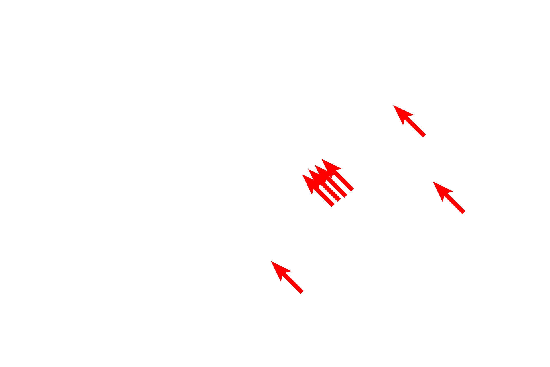
Elastic fibers
An electron micrograph of the reticular layer of the dermis demonstrates large collagen bundles composed of numerous collagen fibrils. Also present are dark elastic fibers criss-crossing the tissue. An active fibroblast is present in the lower part of the image. 5000x
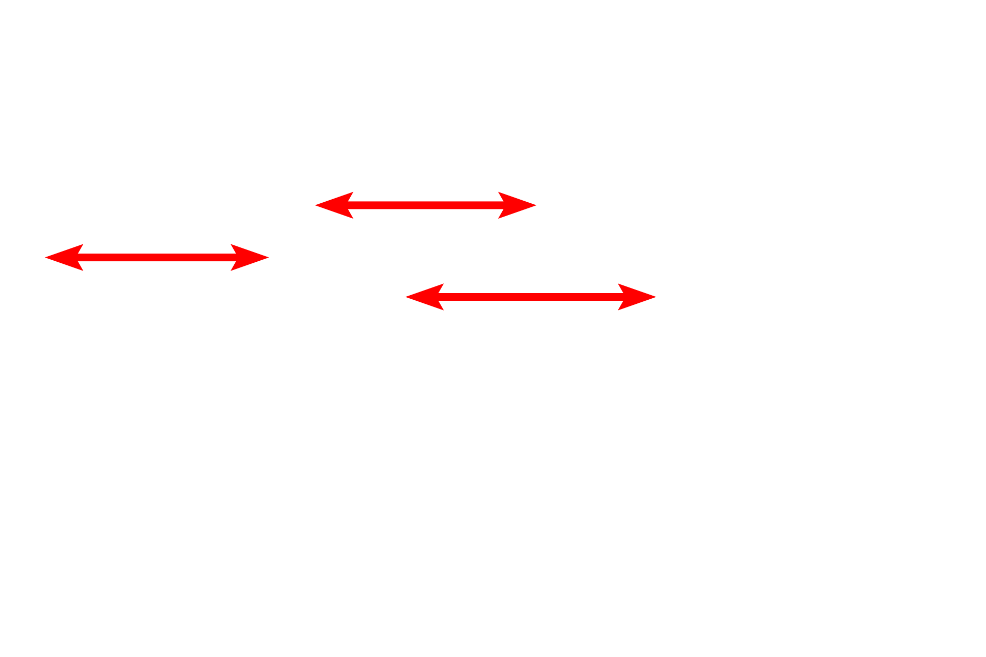
Collagen bundles
An electron micrograph of the reticular layer of the dermis demonstrates large collagen bundles composed of numerous collagen fibrils. Also present are dark elastic fibers criss-crossing the tissue. An active fibroblast is present in the lower part of the image. 5000x
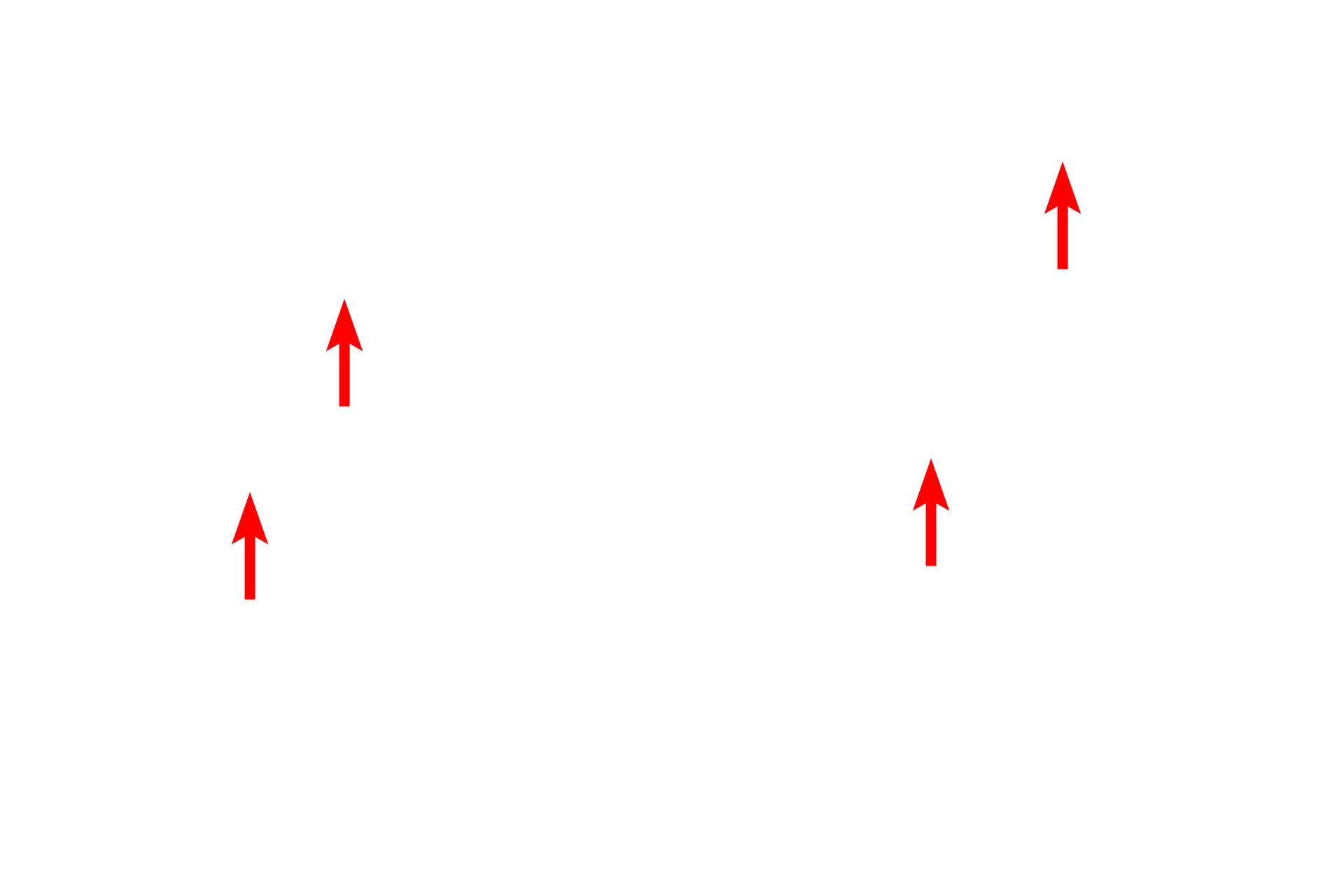
Collagen fibrils
An electron micrograph of the reticular layer of the dermis demonstrates large collagen bundles composed of numerous collagen fibrils. Also present are dark elastic fibers criss-crossing the tissue. An active fibroblast is present in the lower part of the image. 5000x
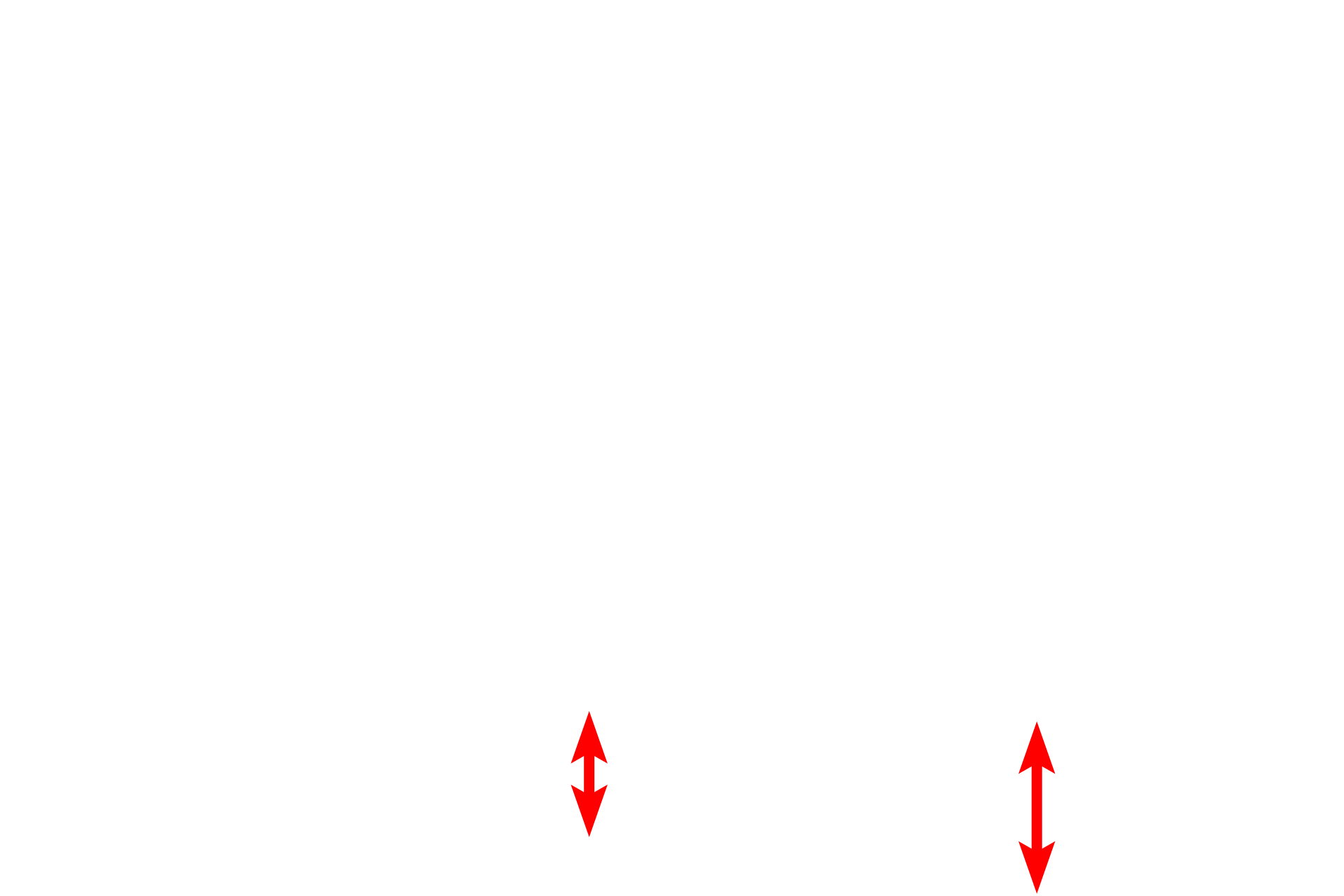
Fibroblast
An electron micrograph of the reticular layer of the dermis demonstrates large collagen bundles composed of numerous collagen fibrils. Also present are dark elastic fibers criss-crossing the tissue. An active fibroblast is present in the lower part of the image. 5000x