
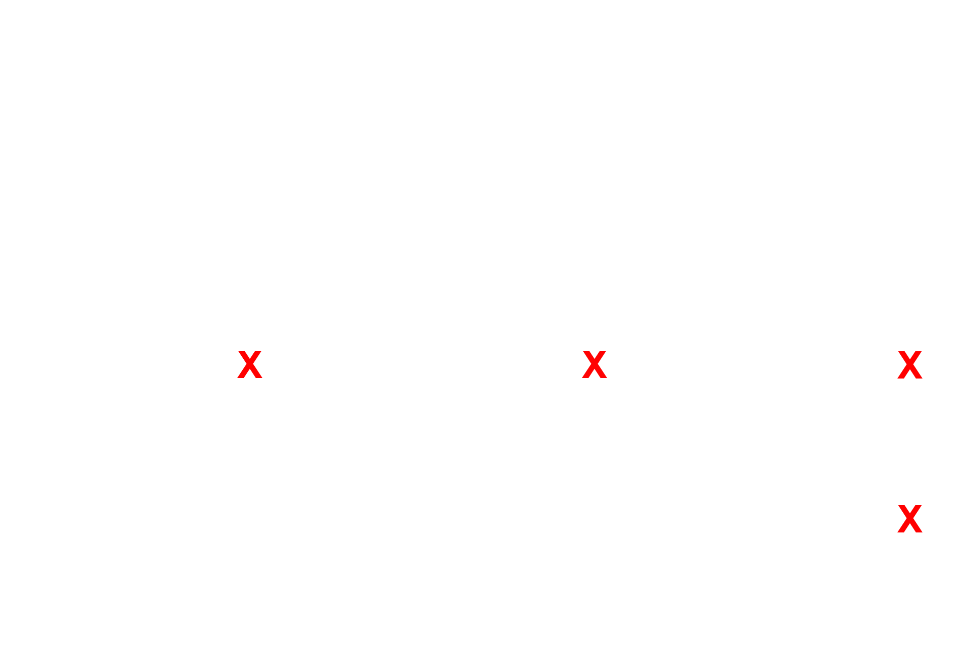
Alveolus
The attenuated cytoplasm of the type I cells lines the interalveolar septum. Septal cells can be identified readily by their vacuoles containing the precursor proteins for surfactant. An alveolar macrophage, lying in in the surfactant layer of the alveolar lumen, is filled with phagocytosed material. 5000x

Alveoli
The attenuated cytoplasm of the type I cells lines the interalveolar septum. Septal cells can be identified readily by their vacuoles containing the precursor proteins for surfactant. An alveolar macrophage, lying in in the surfactant layer of the alveolar lumen, is filled with phagocytosed material. 5000x

Interalveolar septum
The attenuated cytoplasm of the type I cells lines the interalveolar septum. Septal cells can be identified readily by their vacuoles containing the precursor proteins for surfactant. An alveolar macrophage, lying in in the surfactant layer of the alveolar lumen, is filled with phagocytosed material. 5000x

- Type I cells
The attenuated cytoplasm of the type I cells lines the interalveolar septum. Septal cells can be identified readily by their vacuoles containing the precursor proteins for surfactant. An alveolar macrophage, lying in in the surfactant layer of the alveolar lumen, is filled with phagocytosed material. 5000x

- Type II cells
The attenuated cytoplasm of the type I cells lines the interalveolar septum. Septal cells can be identified readily by their vacuoles containing the precursor proteins for surfactant. An alveolar macrophage, lying in in the surfactant layer of the alveolar lumen, is filled with phagocytosed material. 5000x
- Capillaries
The attenuated cytoplasm of the type I cells lines the interalveolar septum. Septal cells can be identified readily by their vacuoles containing the precursor proteins for surfactant. An alveolar macrophage, lying in in the surfactant layer of the alveolar lumen, is filled with phagocytosed material. 5000x

- Endothelial nuclei
The attenuated cytoplasm of the type I cells lines the interalveolar septum. Septal cells can be identified readily by their vacuoles containing the precursor proteins for surfactant. An alveolar macrophage, lying in in the surfactant layer of the alveolar lumen, is filled with phagocytosed material. 5000x
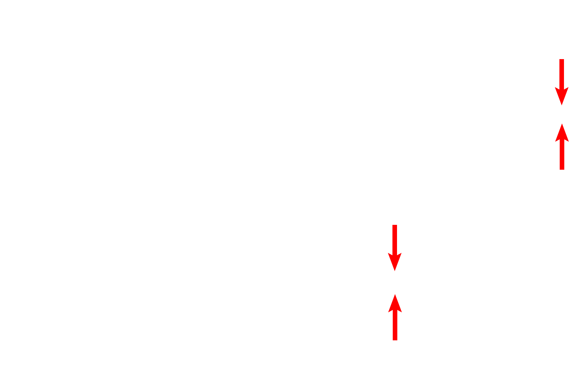
Air-blood barrier
The attenuated cytoplasm of the type I cells lines the interalveolar septum. Septal cells can be identified readily by their vacuoles containing the precursor proteins for surfactant. An alveolar macrophage, lying in in the surfactant layer of the alveolar lumen, is filled with phagocytosed material. 5000x

Macrophage
The attenuated cytoplasm of the type I cells lines the interalveolar septum. Septal cells can be identified readily by their vacuoles containing the precursor proteins for surfactant. An alveolar macrophage, lying in in the surfactant layer of the alveolar lumen, is filled with phagocytosed material. 5000x