
Bronchiole
This higher magnification images shows the components of the bronchiole wall. This larger bronchiole is lined by a pseudostratified columnar epithelium with cilia and goblet cells. Abundant, longitudinally arranged elastic fibers are located beneath the epithelium. The smooth muscle layer is composed of spiraling bands of muscle fibers. 600x

Epithelium
This higher magnification images shows the components of the bronchiole wall. This larger bronchiole is lined by a pseudostratified columnar epithelium with cilia and goblet cells. Abundant, longitudinally arranged elastic fibers are located beneath the epithelium. The smooth muscle layer is composed of spiraling bands of muscle fibers. 600x

- Cilia
This higher magnification images shows the components of the bronchiole wall. This larger bronchiole is lined by a pseudostratified columnar epithelium with cilia and goblet cells. Abundant, longitudinally arranged elastic fibers are located beneath the epithelium. The smooth muscle layer is composed of spiraling bands of muscle fibers. 600x
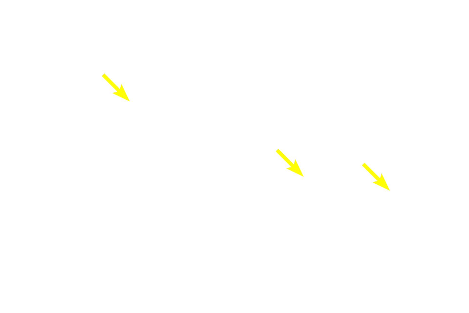
- Goblet cells
This higher magnification images shows the components of the bronchiole wall. This larger bronchiole is lined by a pseudostratified columnar epithelium with cilia and goblet cells. Abundant, longitudinally arranged elastic fibers are located beneath the epithelium. The smooth muscle layer is composed of spiraling bands of muscle fibers. 600x

Basement membrane
This higher magnification images shows the components of the bronchiole wall. This larger bronchiole is lined by a pseudostratified columnar epithelium with cilia and goblet cells. Abundant, longitudinally arranged elastic fibers are located beneath the epithelium. The smooth muscle layer is composed of spiraling bands of muscle fibers. 600x

Elastic fibers
This higher magnification images shows the components of the bronchiole wall. This larger bronchiole is lined by a pseudostratified columnar epithelium with cilia and goblet cells. Abundant, longitudinally arranged elastic fibers are located beneath the epithelium. The smooth muscle layer is composed of spiraling bands of muscle fibers. 600x

Smooth muscle layer
This higher magnification images shows the components of the bronchiole wall. This larger bronchiole is lined by a pseudostratified columnar epithelium with cilia and goblet cells. Abundant, longitudinally arranged elastic fibers are located beneath the epithelium. The smooth muscle layer is composed of spiraling bands of muscle fibers. 600x

Bronchial vessels >
These capillaries are part of the bronchial blood supply that provides oxygen and nutrients to the wall of respiratory passageways. They are not involved in gas exchange with the air.
Image source >
This image was taken of a slide in the University of Virginia collection.